. 2018 Apr 15;200(8):2894-2904.
doi: 10.4049/jimmunol.1701499. Epub 2018 Mar 7.

The TNF Superfamily Molecule LIGHT Promotes the Generation of Circulating and Lung-Resident Memory CD8 T Cells following an Acute Respiratory Virus Infection

Affiliations

The TNF Superfamily Molecule LIGHT Promotes the Generation of Circulating and Lung-Resident Memory CD8 T Cells following an Acute Respiratory Virus Infection

Pritesh Desai et al. J Immunol. .

Abstract

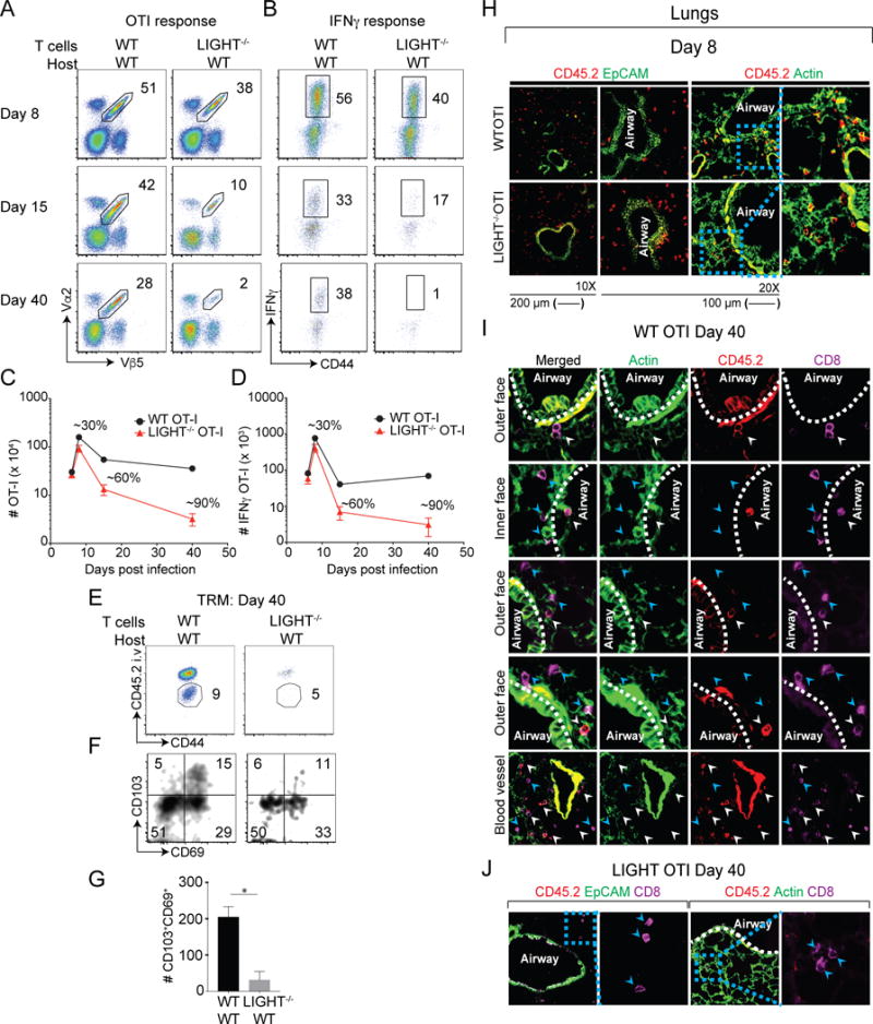
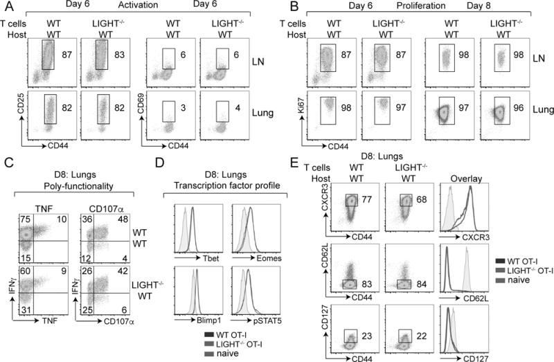
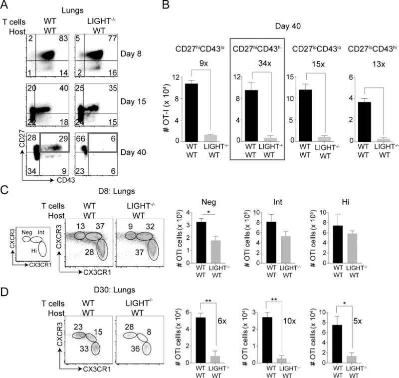
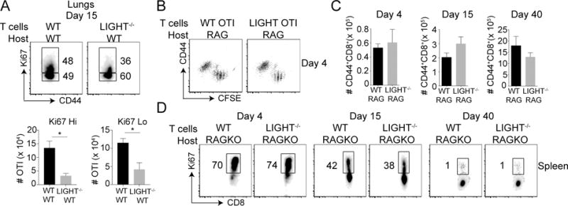
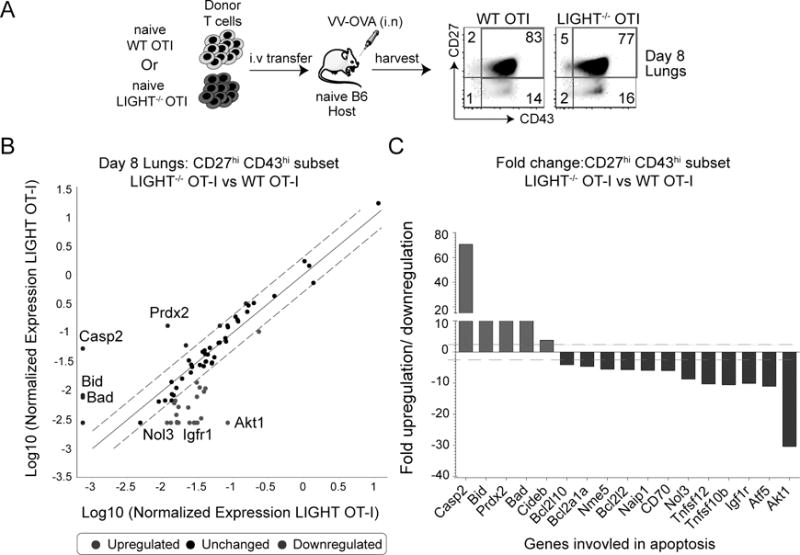
The transition of effector T cells or memory precursors into distinct long-lived memory T cell subsets is not well understood. Although many molecules made by APCs can contribute to clonal expansion and effector cell differentiation, it is not clear if clonal contraction and memory development is passive or active. Using respiratory virus infection, we found that CD8 T cells that cannot express the TNF family molecule lymphotoxin-like, exhibits inducible expression, competes with HSV glycoprotein D for herpes virus entry mediator, a receptor expressed by T lymphocytes (LIGHT) are unimpaired in their initial response and clonally expand to form effector cell pools. Thereafter, LIGHT-deficient CD8 T cells undergo strikingly enhanced clonal contraction with resultant compromised accumulation of both circulating and tissue-resident memory cells. LIGHT expression at the peak of the effector response regulates the balance of several pro- and antiapoptotic genes, including Akt, and has a preferential impact on the development of the peripheral memory population. These results underscore the importance of LIGHT activity in programming memory CD8 T cell development, and suggest that CD8 effector T cells can dictate their own fate into becoming memory cells by expressing LIGHT.


Figures

Figure 1
Figure 1. LIGHT expressed by CD8 T cells regulates formation of memory cells
Equal numbers (5 × 10⁴) of WT and LIGHT−/− naïve (CD44lo) OT-I (Vα2+Vβ5+) transgenic CD8 T cells were adoptively transferred into BL/6 mice and infected with rVacV-WR-OVA (2 × 10⁴ PFU i.n) the following day. (A-D) Lungs were harvested at days 6, 8, 15 and 40 post-infection and stained for (A) CD8, CD44, Vα2, and Vβ5 and frequencies of OT-I CD8 T cells determined. (C) OT-I cells from lungs of recipients were re-stimulated in vitro with OVA peptide and stained intra-cellularly with IFN-γ. Absolute numbers of (C) OT-I cells and (D) IFNγ+ OT-I cells from lungs were quantified post-infection. (E-G) At day 40 post-infection, recipient mice were injected with anti-CD45.2 antibody intravenously, three minutes before euthanizing. (H-J) Equal numbers (5 × 10⁵) of WT and LIGHT−/− naïve (CD44lo) OT-I (Vα2+Vβ5+) transgenic CD45.2+CD8 T cells were adoptively transferred into CD45.1+SJL mice and infected with rVacV-WR-OVA (2 × 10⁴ PFU i.n) the following day. IF analysis of frozen (H) lung sections from recipient mice stained for CD45.2 (red) and CD8 (magenta) at day 8 post-infection. Similar analysis for was done at day 40 post-infection in (I & J) the lungs. ActinGreen or EpCAM (green) were used to visualize lung morphology. Bars, 200 μm & 100 μm respectively. Similar results were obtained in two independent experiments. Representative plots of CD45.2 and CD44 were pre-gated on CD8+OT-I (Vα2+Vβ5+) cells. The gated CD45.2 negative cells were analyzed for the expression of CD103 and CD69 using flow cytometry and total numbers of cells calculated in lungs. *, P<0.05; and results are the mean ± SEM (n = 3 mice/group). Similar results were obtained in four independent experiments.
Figure 2
Figure 2. Intact early activation, proliferation, poly-functionality and differentiation of CD8 T cells in the absence of LIGHT
Mice that received WT or LIGHT−/− CD8 T cells in Fig 1 were analyzed on day 6 and 8 post-infection. (A-E) Lung cells were stained extracellularly with antibodies to CD8, Vα2, Vβ5, CD44, CD25, CD69, CXCR3, CD62L, IL7Rα (CD127); (B) intranuclearly with Ki67; (C) intracellularly with antibodies to TNF, IFNγ following re-stimulation with SIINFEKL peptide during which CD107α was added; and (D) intra-nuclearly with antibodies to t-bet, eomes, blimp1, pStat5, without peptide re-stimulation.
Figure 3
Figure 3. Differential impact of a LIGHT deficiency on memory CD8 T cell subsets
(A) Mice that received WT or LIGHT−/− CD8 T cells as in Fig 1 were analyzed on day 8, 15 and 40 post-infection. Lung cells were stained with extracellular antibodies for CD8, Vα2, Vβ5, CD44, CD27, CD43. (B) Absolute cell numbers of CD27 vs. CD43 subsets at day 40 post-infection were quantified. Similar results were obtained in three independent experiments and results are the mean ± SEM (n = 3 mice/group). (C-D) Lungs of WT or LIGHT−/− CD8 T cell recipient mice were harvested at (C) day 8 and (D) day 30 post-infection and stained for CD8, Vα2, Vβ5, CD44, CXCR3 and CX3CR1 and corresponding cell numbers of subsets were quantified. Single experiment with 4 mice in a group and results are the mean ± SEM. Statistical significance was performed using student t test with * >0.05 and ** >0.005.
Figure 4
Figure 4. Intact homeostatic proliferation in the absence of LIGHT
(A) Lung cells from mice that received WT or LIGHT−/− CD8 T cells were analyzed on day 15 post-infection with VacV as in Fig. 1. After staining cells for CD8, Vα2, Vβ5, and CD44, cells were stained intra-nuclearly with Ki67 (B-D) Equal numbers (5 × 10⁵) of WT and LIGHT−/− naïve (CD44lo) OT-I (Vα2+Vβ5+) transgenic CD8 T cells were CFSE labeled and adoptively transferred into RAG-deficient mice without infection. Spleens were harvested from recipient mice at day 4, 15 and 40 post-infection and cells were stained extracellularly for CD8, Vα2, Vβ5, CD44, and intranuclearly with Ki67. *, P<0.05. Similar results were obtained in two independent experiments and results are the mean ± SEM (n = 3 mice/group).
Figure 5
Figure 5. LIGHT regulates the survival of effector CD8 T cells
(A) Lungs from mice that received WT or LIGHT−/− CD8 T cells were harvested at day 8 post-infection with VacV and stained for CD8, Vα2, Vβ5, CD44, CD27 and CD43. The CD27hiCD43hi subset was sort purified and total mRNA was isolated. (B) Transcript levels of apoptotic genes were measured using affymetrix mouse apoptotic gene arrays and presented as a scatter plot with dotted lines representing 2-fold differences between the two groups. (C) Bars represent fold change in transcript levels of the CD27hiCD43hi subset between LIGHT-deficient CD8 T cells and WT CD8 T cells. Inset: Lung cells from WT and LIGHT−/− OT-I recipient mice were stained intra-nuclearly with pAKT.
Figure 6
Figure 6. Endogenous HVEM acts as a ligand for LIGHT made by CD8 T cells
(A & B) Equal numbers (5 × 10⁴) of WT CD8 T cells were adoptively transferred into naive WT and HVEM−/− mice, and compared to LIGHT−/− CD8 T cells transferred into WT mice. All recipient mice were infected with rVacV-WR-OVA (2 × 10⁴ PFU i.n) the following day. At day 40 post-infection, lung and spleen cells were stained for CD8, Vα2, Vβ5, and CD44 and analyzed by flow cytometry. *, P<0.05, **, P<0.01. Similar results were obtained in two independent experiments and results are the mean ± SEM (n = 3 mice/group). (C) Lungs and mediastinal lymph nodes from WT CD8 T cell recipient mice were harvested on day 0 (naïve), 1, 3 and 12 post-infection. Dendritic cells and various innate cells were stained for HVEM. Grey histogram represents cells from HVEM−/− mice. (D) Lung cells were incubated in vitro with either recombinant VV-S-OVA-GFP or VV-WR at MOI of 1. Six-hours post-incubation, cells were stained with antibodies specific for lung DC subsets and inflammatory monocytes and 25D1 that recognizes S-OVA-MHC-I complexes.
Figure 7
Figure 7. LIGHT controls memory and recall responses to influenza virus infection
(A) Equal numbers (5 × 10⁴) of WT and LIGHT−/− naïve (CD44lo) OT-I (Vα2+Vβ5+) transgenic CD8 T cells were adoptively transferred into WT mice and infected with PR8-SIINFEKL (i.n) the following day. Lungs were harvested at day 40 post-infection and stained for CD8, CD44, Vα2, Vβ5 and NP-tetramer (top and bottom). OT-I cells from lungs of recipients were re-stimulated in vitro with OVA peptide and stained intra-cellularly with IFN-γ (middle). (B & C) WT & LIGHT−/− CD8 T cell recipient mice that were previously infected with rVacV-WR-OVA (i.n) were re-infected with PR8-SIINFEKL (i.n) after 40 days from primary infection. Lungs were harvested at day 6 and stained for CD8, CD44, Vα2, Vβ5 and NP-tetramer. (D) Numbers and fold change in the cell numbers between WT CD8 T cells and LIGHT−/− CD8 T cells from day 40 post-primary infection to day 6 post re-infection was quantified. *, P<0.05. Similar results were obtained in two independent experiments and results are the mean ± SEM (n = 3 mice/group).

References

    1. Woodland DL, Kohlmeier JE. Migration, maintenance and recall of memory T cells in peripheral tissues. Nat Rev Immunol. 2009;9:153–161. - PubMed
    1. Lauvau G, Boutet M, Williams TM, Chin SS, Chorro L. Memory CD8(+) T Cells: Innate-Like Sensors and Orchestrators of Protection. Trends Immunol. 2016;37:375–385. - PMC - PubMed
    1. Sallusto F, Lenig D, Forster R, Lipp M, Lanzavecchia A. Two subsets of memory T lymphocytes with distinct homing potentials and effector functions. Nature. 1999;401:708–712. - PubMed
    1. Lanzavecchia A, Sallusto F. Dynamics of T lymphocyte responses: intermediates, effectors, and memory cells. Science. 2000;290:92–97. - PubMed
    1. Wherry EJ, Teichgraber V, Becker TC, Masopust D, Kaech SM, Antia R, von Andrian UH, Ahmed R. Lineage relationship and protective immunity of memory CD8 T cell subsets. Nat Immunol. 2003;4:225–234. - PubMed

Publication types

MeSH terms

Substances