An official website of the United States government
The .gov means it’s official.
Federal government websites often end in .gov or .mil. Before
sharing sensitive information, make sure you’re on a federal
government site.
The site is secure.
The https:// ensures that you are connecting to the
official website and that any information you provide is encrypted
and transmitted securely.
The essential histone chaperone FACT (facilitates chromatin transcription) promotes both nucleosome assembly and disassembly. FACT is a heterodimer of Spt16 with either SSRP1 or Pob3, differing primarily by the presence of a high-mobility group B (HMGB) DNA-binding domain furnished only by SSRP1. Yeast FACT lacks the intrinsic HMGB domain found in SSRP1-based homologs such as human FACT, but yeast FACT activity is supported by Nhp6, which is a freestanding, single HMGB-domain protein. The importance of histone binding by FACT domains has been established, but the roles of DNA-binding activity remain poorly understood. Here, we examined these roles by fusing single or multiple HMGB modules to Pob3 to mimic SSRP1 or to test the effects of extended DNA-binding capacity. Human FACT and a yeast mimic both required Nhp6 to support nucleosome reorganization in vitro, indicating that a single intrinsic DNA-binding HMGB module is insufficient for full FACT activity. Three fused HMGB modules supported activity without Nhp6 assistance, but this FACT variant did not efficiently release from nucleosomes and was toxic in vivo Notably, intrinsic DNA-binding HMGB modules reduced the DNA accessibility and histone H2A-H2B dimer loss normally associated with nucleosome reorganization. We propose that DNA bending by HMGB domains promotes nucleosome destabilization and reorganization by exposing FACT's histone-binding sites, but DNA bending also produces DNA curvature needed to accommodate nucleosome assembly. Intrinsic DNA-bending activity therefore favors nucleosome assembly by FACT over nucleosome reorganization, but excessive activity impairs FACT release, suggesting a quality control checkpoint during nucleosome assembly.
The authors declare that they have no conflicts of interest with the contents of this article
Figures
Figure 1.
Architecture of Pob3/SSRP1 constructs used.…
Figure 1.
Architecture of Pob3/SSRP1 constructs used. Native Pob3 and Nhp6 proteins ( S. cerevisiae…
Figure 1.
Architecture of Pob3/SSRP1 constructs used. Native Pob3 and Nhp6 proteins (S. cerevisiae) are drawn to scale, with the N-terminal (Pob3-N (light green)), middle (Pob3-M (dark green)), and acidic domains (AD (red)) of Pob3 and the HMGB domains (blue) of various proteins indicated as ovals flanked by unstructured linkers. Similar domains found in human SSRP1 are shown with the same color scheme but with orange outlines. SAB indicates the serine-acidic-basic domain of hSSRP1. The native architecture of human HMGB1 is shown to contrast with the single HMGB motif architecture of Nhp6. Various fusion proteins with combinations of these domains used here are shown below the gray line.
Figure 2.
Purification of factors and consequences…
Figure 2.
Purification of factors and consequences of their expression in vivo . A ,…
Figure 2.
Purification of factors and consequences of their expression in vivo.A, 2.5 μg of purified SP (Spt16–Pob3) or the derivatives with one or three Nhp6 modules fused to Pob3 (-1N, -3N) were separated by SDS-PAGE and stained with Coomassie Blue dye. B, top panel, strain 7789-2-1 pJW4 (Table S1, pob3-Δ YEp URA3 POB3) was transformed with pTF162 (YEp LEU2 vector), pTF139 (YCp LEU2 POB3 (16)), pHX13 (YEp LEU2 GAL1p-POB3) or pHX16 (YEp LEU2 GAL1p-POB3–3N). Cultures were grown in rich medium to permit loss of pJW4, and then 10-fold serial dilutions were placed on synthetic glucose medium lacking leucine (Glc −leu) or containing fluoroorotic acid (Glc FOA) to determine retention of the LEU2 plasmids and loss of the URA3 plasmid, respectively (50). Complementation of pob3-Δ was robust for the YCp POB3 plasmid, weaker for the YEp POB3 plasmid under repressive conditions for the GAL1 promoter, and absent for the YEp POB3–3N plasmid. Expression of POB3 in this configuration was therefore adequate to produce some growth, but Pob3–3N either did not complement pob3-Δ or its toxicity outweighed any benefit. Bottom panel, strain 8127-7-4 (WT, Table S1) was transformed with the same plasmids as above and tested under selection for the plasmids on medium that repressed (Glc) or induced (Gal) the GAL1 promoter. GAL1p-POB3–3N blocked growth only on Gal, demonstrating toxicity of the Pob3–3N protein.
Figure 3.
Analysis of complex formation between…
Figure 3.
Analysis of complex formation between SP, SP–1N, SP–3N, or hFACT and DNA or…
Figure 3.
Analysis of complex formation between SP, SP–1N, SP–3N, or hFACT and DNA or nucleosomes.A, Spt16–Pob3 (SP) or the SP–1N derivative were mixed at the indicated concentrations with a 181-bp DNA fragment, and then the fraction of total bound DNA was determined following native PAGE (Fig. S1). The average ± S.D. is shown for four independent repeats. B, same as in A, except Nhp6A and hFACT were tested, and the results from three repeats are shown. Subsequent assays were performed with 200 nm hFACT, a condition that demonstrated little or no binding to free DNA in this assay. C, nucleosomes were mixed with 200 nm SP or SP–1N and the indicated concentrations of Nhp6, and the bound fraction (FACT:Nuc complexes as shown in D) was determined following native PAGE. The average ± S.D. for four independent repeats is shown. D, nucleosomes alone or mixed with 200 nm SP, SP–1N, or SP–3N without or with 2 μm Nhp6 were separated by native PAGE. Gels were scanned to detect the Cy5-labeled DNA (green signal) and the Oregon Green-labeled H2B (red signal) with the merged images on the right. Complexes formed between FACT and displaced dimers are indicated as SP-x:H2A-H2B, and a typically weak band whose prominence increased in proportion to the number of Nhp6 modules fused to Pob3 is indicated as a Novel Complex.
Figure 4.
Effect of fused Nhp6 modules…
Figure 4.
Effect of fused Nhp6 modules on nuclease sensitivity. A , 200 n m…
Figure 4.
Effect of fused Nhp6 modules on nuclease sensitivity.A, 200 nm SP or SP–1N was mixed with nucleosomes, and then DraI was added and the amount of digestion determined after 8, 16, and 24 min by denaturing SDS-PAGE analysis. The initial rate of digestion was determined from the time courses and normalized to the value with SP + 2 μm Nhp6 tested in parallel. A similar experiment with three repeats gave no digestion for SP and SP–1N without Nhp6, and the rate for SP–1N + N was 68% ± 5% of the SP + N rate (not shown). B, same as in A, except the concentration of SP–3N was varied with or without 2 μm Nhp6 and the digestion rate was compared with a parallel control reaction with 200 nm SP and 2 μm Nhp6. C, same as in B, except four repeats were performed with 20 nm and 200 nm SP–3N with the average ± S.D. of the absolute rate of digestion (% of total DNA digested/min) for each condition reported without normalization. D, similar to B, except an AT-hook nucleosome positioning sequence was used (4) and the rate of digestion with PstI was measured.
Figure 5.
Fusion of Nhp6 to Pob3…
Figure 5.
Fusion of Nhp6 to Pob3 inhibits H2A–H2B displacement from nucleosomes by FACT. A…
Figure 5.
Fusion of Nhp6 to Pob3 inhibits H2A–H2B displacement from nucleosomes by FACT.A, complexes were formed as described in the legend for Fig. 3D. The H2A–H2B content in the FACT:Nuc band (see Fig. 3D) was determined by comparing the ratio of dimer signal to DNA signal and normalizing it to intact nucleosomes. B, samples were prepared and analyzed as described in the legend for Fig. 3D, except competitor DNA was added prior to electrophoresis. C, the H2A–H2B content of the released nucleosomes prepared as in B above was determined as in A. The average ± S.D. for four independent repeats is shown. D, the fraction of signal remaining in the Retained Complexes region defined in B was calculated for the same samples as described for C.
Figure 6.
Effects of Pob3–1N in vivo…
Figure 6.
Effects of Pob3–1N in vivo and characterization of Nhp6 levels. A , 10-fold…
Figure 6.
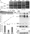
Effects of Pob3–1N in vivo and characterization of Nhp6 levels.A, 10-fold serial dilutions of strains with the genotypes indicated (Table S1) were tested as indicated. YPAD is rich medium (yeast extract, peptone, dextrose, adenine sulfate), HU150 is YPAD with 150 mm hydroxyurea, SC Glc is synthetic complete medium with glucose as the carbon source, and −Lys is the same but lacking lysine. Growth on −Lys reveals the Spt− phenotype in these strains with the lys2–128∂ allele (46). B, purified recombinant Nhp6A or 7 μg of extract from cells with the genotypes shown (Table S1) were separated by SDS-PAGE, transferred to nitrocellulose, and then detected with polyclonal antiserum directed against Nhp6A protein. The signal from the western blotting was quantitated with NIH ImageJ software (51), yielding the standard curve in C. Total amounts of Nhp6 were calculated from the standard curve and converted to copy number/cell (in D, the average ± S.D. for eight independent repeats is shown) using the previously determined relationship between cell number and total protein in these extracts (49). E, same as in B, except strains lacking Nhp6 and with Pob3–Nhp6 were included. The top panel shows a short exposure to optimize detection of Pob3–1N with Nhp6 antiserum, the middle panel shows the entire gel at a medium exposure time, and the bottom panel shows a long exposure time for the region containing free Nhp6 to emphasize the lack of signal in nhp6-ΔΔ strains. F, same as in E, except the gel was probed with antibodies against Pob3. A cross-reacting species observed in all strains but visible as a distinct band only with Pob3–1N fusions is marked by an asterisk.
Figure 7.
Human FACT reversibly reorganizes nucleosomes…
Figure 7.
Human FACT reversibly reorganizes nucleosomes in the presence of added Nhp6. A ,…
Figure 7.
Human FACT reversibly reorganizes nucleosomes in the presence of added Nhp6.A, nucleosomes with SP or human FACT with or without 2 μm Nhp6 were separated by native PAGE as described in the legend for Fig. 3. B, complexes were treated with DraI as described in the legend for Fig. 4, except a single 10-min time point was analyzed for three independent samples of 200 nm SP, 2 μm Nhp6 (yFACT + N) or 200 nm hFACT, 2 μm Nhp6 (hFACT + N) with the average ± S.D. shown. The absolute rate of digestion varies with batches of DraI (compare with Fig. 4C). No digestion was observed in parallel experiments with hFACT, SP, or Nhp6 alone (not shown). C, FACT–nucleosome complexes formed as in A were treated with competitor DNA, and the H2A–H2B dimer content in the resulting dissociated nucleosomes was determined as described in the legend for Fig. 5C, with the average ± S.D. of three independent samples shown. D, spFRET analysis of nucleosome N35/112 (Nuc) alone and then with hFACT + Nhp6 before and after the addition of competitor DNA (+DNA) is shown (see Refs. and for details and additional control experiments). Frequency distributions of proximity ratios (EPR) calculated from the FRET efficiencies of individual nucleosomes were based on analysis of 4,315 (Nuc), 16,233 (hFACT + Nhp6), and 5,141 (hFACT + Nhp6 + competitor DNA) single particles. Control experiments published previously show that the Cy3 and Cy5 fluorophores inserted 35 and 112 bp into this 603-nucleosome positioning sequence produced high EPR values only when the DNA was tightly coiled around a histone core (10). Neither hFACT nor Nhp6 alone altered the EPR distribution in this assay (10, 11), but their combination caused a dramatic decrease in EPR values (hFACT + Nhp6). This effect was reversible, as the addition of competitor DNA restored the characteristic EPR distribution for nucleosomes (hFACT + Nhp6 + competitor DNA). Human FACT therefore caused the same disruption of nucleosome structure (uncoiling of DNA that affects about 70% of the DNA in a nucleosome) that was observed for yeast FACT (10). This depended on the further addition of HMGB proteins beyond the single domain available in SSRP1 and was reversible. Inset, an approximate representation of N35/112.
Figure 8.
Model for the role of…
Figure 8.
Model for the role of the DNA-binding module in FACT activity. FACT makes…
Figure 8.
Model for the role of the DNA-binding module in FACT activity. FACT makes multiple weak contacts with a nucleosome leading to a more permissive but unstable “stressed” form. Converting this to a fully reorganized form requires multiple DNA-binding events to sequentially expose higher-affinity binding sites, allowing the formation of a stable complex detectable by EMSA. This activity can be provided by high concentrations of free Nhp6 or by multiple fused HMGB domains on Pob3 but not by the single HMGB domain in SSRP1 or Pob3–1N. Resolution of the reorganized form requires DNA-bending activity to produce curved DNA to enclose the H2A–H2B dimers, and an intrinsic HMGB domain favors this, skewing the equilibrium toward form 2. This wrapping step has similarities with the proposed role of the ATP-dependent remodeler ACF in converting pre-nucleosomes to nucleosomes (48), suggesting potential parallels between active and passive nucleosome assembly processes. The DNA in form 2 is tightly coiled, producing the same spFRET and nuclease sensitivity profiles as canonical nucleosomes (11), but this form is not fully resolved and can promote active processes like transcription or remodeling. Complete resolution requires coordinated assessment of the integrity of the nucleosome, with FACT variants like SP–3N (Fig. 5) and Spt16–Pob3–Q308K failing to release efficiently due to inefficient completion of this checkpoint (42). Factors are drawn approximately to scale using the color scheme as described in Fig. 1 and the following Protein Data Bank structures: 1ID3 (52), 1J5N (25), 4IOY (53), 2GCL (47), 3BIT (54), 4KHB (55), 4WNN, (36) and 4Z2M (3).
Tsunaka Y, Ohtomo H, Morikawa K, Nishimura Y.Tsunaka Y, et al.iScience. 2020 Oct 6;23(10):101641. doi: 10.1016/j.isci.2020.101641. eCollection 2020 Oct 23.iScience. 2020.PMID: 33103079Free PMC article.
Formosa T. (2012) The role of FACT in making and breaking nucleosomes. Biochim. Biophys. Acta 1819, 247–255 10.1016/j.bbagrm.2011.07.009
-
DOI
-
PMC
-
PubMed
Hondele M., and Ladurner A. G. (2013) Catch me if you can: How the histone chaperone FACT capitalizes on nucleosome breathing. Nucleus 4, 443–449 10.4161/nucl.27235
-
DOI
-
PMC
-
PubMed
Tsunaka Y., Fujiwara Y., Oyama T., Hirose S., and Morikawa K. (2016) Integrated molecular mechanism directing nucleosome reorganization by human FACT. Genes Dev. 30, 673–686 10.1101/gad.274183.115
-
DOI
-
PMC
-
PubMed
Xin H., Takahata S., Blanksma M., McCullough L., Stillman D. J., and Formosa T. (2009) yFACT induces global accessibility of nucleosomal DNA without H2A-H2B displacement. Mol. Cell 35, 365–376 10.1016/j.molcel.2009.06.024
-
DOI
-
PMC
-
PubMed
Rhoades A. R., Ruone S., and Formosa T. (2004) Structural features of nucleosomes reorganized by yeast FACT and its HMG box component, Nhp6. Mol. Cell. Biol. 24, 3907–3917 10.1128/MCB.24.9.3907-3917.2004
-
DOI
-
PMC
-
PubMed