. 2018 Apr 20;293(16):5781-5792.
doi: 10.1074/jbc.RA117.001289. Epub 2018 Mar 7.

Structural basis for the regulatory interaction of the methylglyoxal synthase MgsA with the carbon flux regulator Crh in Bacillus subtilis

Affiliations

Structural basis for the regulatory interaction of the methylglyoxal synthase MgsA with the carbon flux regulator Crh in Bacillus subtilis

Achim Dickmanns et al. J Biol Chem. .

Abstract

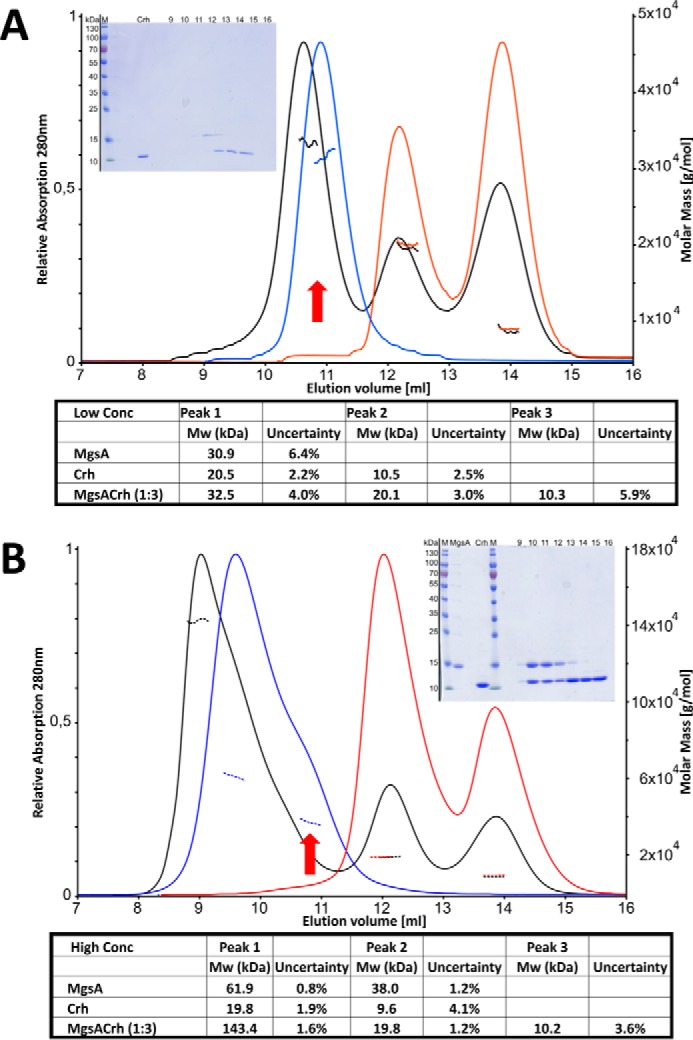
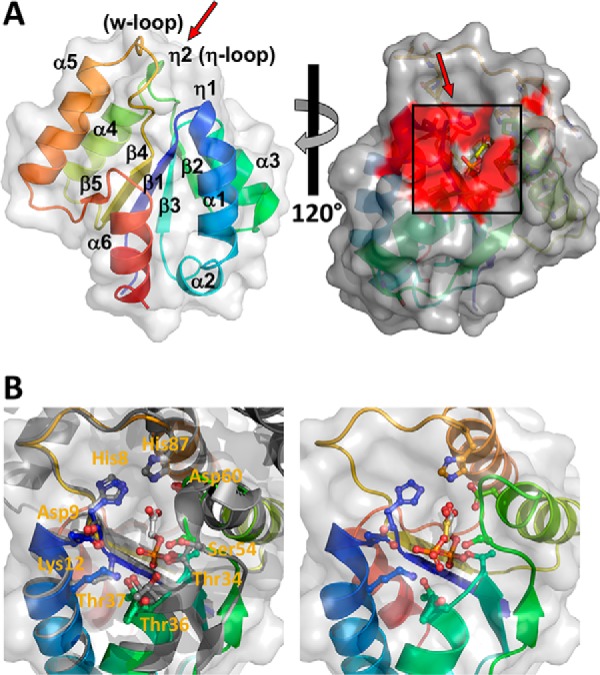
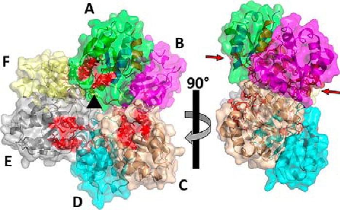
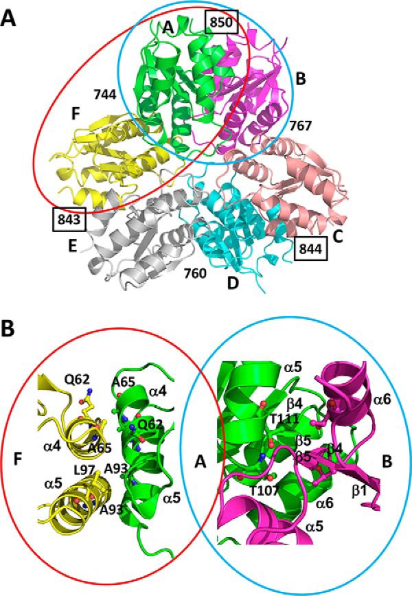
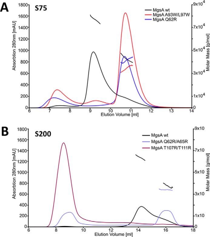
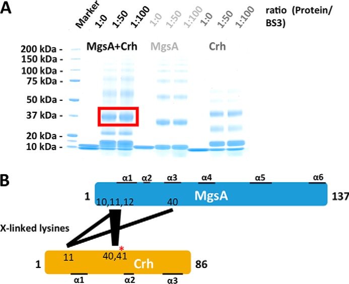
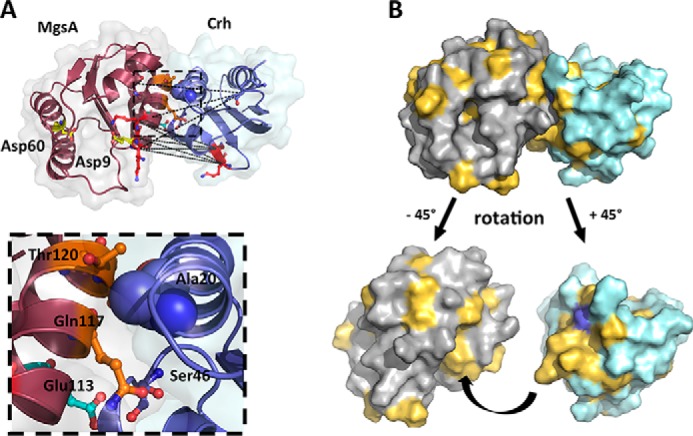
Utilization of energy-rich carbon sources such as glucose is fundamental to the evolutionary success of bacteria. Glucose can be catabolized via glycolysis for feeding the intermediary metabolism. The methylglyoxal synthase MgsA produces methylglyoxal from the glycolytic intermediate dihydroxyacetone phosphate. Methylglyoxal is toxic, requiring stringent regulation of MgsA activity. In the Gram-positive bacterium Bacillus subtilis, an interaction with the phosphoprotein Crh controls MgsA activity. In the absence of preferred carbon sources, Crh is present in the nonphosphorylated state and binds to and thereby inhibits MgsA. To better understand the mechanism of regulation of MgsA, here we performed biochemical and structural analyses of B. subtilis MgsA and of its interaction with Crh. Our results indicated that MgsA forms a hexamer (i.e. a trimer of dimers) in the crystal structure, whereas it seems to exist in an equilibrium between a dimer and hexamer in solution. In the hexamer, two alternative dimers could be distinguished, but only one appeared to prevail in solution. Further analysis strongly suggested that the hexamer is the biologically active form. In vitro cross-linking studies revealed that Crh interacts with the N-terminal helices of MgsA and that the Crh-MgsA binding inactivates MgsA by distorting and thereby blocking its active site. In summary, our results indicate that dimeric and hexameric MgsA species exist in an equilibrium in solution, that the hexameric species is the active form, and that binding to Crh deforms and blocks the active site in MgsA.

Keywords: Bacillus; Crh; HPr; bacteria; bacterial genetics; bacterial metabolism; catabolite regulation; crystal structure; glucose catabolism; glycolysis; metabolic regulation; methylglyoxal synthase; methylglyoxal toxicity; phosphoprotein; prokaryotic signal-transduction; protein cross-linking; protein structure; protein-protein interaction.


Conflict of interest statement

The authors declare that they have no conflicts of interest with the contents of this article

Figures

Figure 1.
Figure 1.
The MgsA binds to Crh only if MgsA is present in hexamers. SEC-MALS experiments suggest that only the MgsA hexamer binds Crh, resulting in a 1:1 stoichiometry. A, low MgsA concentration (13 μm). B, high MgsA concentration (135 μm). In each case, Crh was used in a 3-fold molar excess. Insets, Coomassie-stained gel of the respective fractions (equivalent to the ml of elution volume) obtained in the SEC run of the MgsA/Crh mixture. Red arrows, elution volume of dimeric MgsA. Below each chromatogram, the calculated molar masses determined by MALS are listed. Blue lines, MgsA; red lines, Crh; black lines, mixture of MgsA and Crh (concentrations as indicated). Dotted line, molecular masses as determined by MALS.
Figure 2.
Figure 2.
The structure of B. subtilis MgsA exhibits an overall globular shape. A, the central five-stranded β-sheet is flanked by six α-helices. The coloring is in a rainbow from the N terminus (blue) to the C terminus (red). The surface structure is depicted in gray; the individual secondary structure motifs are indicated and numbered as α-helices (α) or β-strand (β) from the N to the C terminus. The panel on the right shows a surface representation of an MgsA monomer in gray with the surface region of important active site residues highlighted in red. B, left, magnification of the active site as deduced from an overlay with the E. coli MgsA (gray) with 2-phosphoglycolic acid (ball-and-stick mode, PDB code 1EGH) bound, serving as reference ligand. Cavity conformation and arrangement of active site residues are highly conserved (see also Figs. S3 and S5). Identical residues of B. subtilis MgsA within the Mgs family involved in ligand binding are depicted with their side chains in ball-and-stick mode with the carbon atoms labeled according to A, with oxygen in red and nitrogen in blue. Residue numbering is according to B. subtilis MgsA. Right, result of a docking experiment with the substrate dihydroxyacetone phosphate (carbons in yellow, phosphorus in orange) and phosphoglycolohydroxamic acid (PDB code 1IK4) as reference.
Figure 3.
Figure 3.
Structure of the B. subtilis MgsA hexamer. The hexamer is composed of three dimers of MgsA, which arrange in the hexamer with three active sites (active-site residues indicated in red) arranged by 120° rotations (indicated by the black triangle in the center) on either side of the hexamer. The individual molecules are indicated in different colors and labeled A–F (top right panel). Bottom right panel, side view of the hexamer. The arrows indicate the active sites of molecules A and B.
Figure 4.
Figure 4.
Mutational and structural analysis suggests hexamer disassembly into MgsA dimers. A, the disassembly of the hexamer (molecules colored and labeled as in Fig. 3) into dimers could result in two different dimers, one leaving the active sites of one subunit in contact with the neighboring molecule (AF, indicated by a red ellipsoid) or disrupting the active site connecting surface (AB, indicated by a cyan ellipsoid). The numbers depicted indicate the average surface area (in Å2) of the individual molecules involved in interaction with the neighboring molecule. See “The functional MgsA dimer” for details. B, magnification of the interfaces AF and AB. The point mutations tested for interference with hexamer formation of MgsA are indicated, and only those located at the interface formed by the MgsA molecules A and F prevent hexamerization. Thus, the monomers that interact to form the stable dimer require the other dimer interface (e.g. AB).
Figure 5.
Figure 5.
The analysis of the different MgsA variants suggests that the AB-type dimer is the one that exists in solution not the AF-type dimer. SEC-MALS experiments suggest that only the WT MgsA is folded correctly and capable of forming hexamers. The solid lines indicate the absorption profile of the SEC run, whereas the stray lines indicate the molecular mass observed in the MALS measurement at the respective elution volume. A, in contrast to WT MgsA, the point mutants A93W/L97W and Q69R form predominantly soluble dimers. B, opposing the WT MgsA, the point mutant Q62R/A65R forms mostly dimers and T107R/T111R aggregates. See “The functional MgsA dimer” for details.
Figure 6.
Figure 6.
Determination of Crh cross-links to MgsA. A, an in vitro chemical cross-linking of MgsA and Crh either alone or in a complex formed in a 1:1 ratio. The samples were treated with the predetermined (Fig. S8) optimal amounts of BS3 using a 1:50 and 1:100 protein/cross-linker ratio. According to the size, bands that represent the complex of MgsA and/or MgsA-Crh were analyzed by LC-MS. B, schematic representation of the intermolecular cross-links determined (see also Table 3 and Figs. S5 and S6).
Figure 7.
Figure 7.
Model of the MgsA-Crh interaction. A, the model of MgsA-Crh complex obtained by blind docking experiments fulfills the described criteria. The MgsA molecule (gray surface and molecule colored dark red) is in close contact with Crh (surface in light cyan and the molecule colored in purple-blue) in the vicinity of the MgsA active site. For reference, the catalytically active site residues of MgsA, Asp9 and Asp60, are depicted in ball-and-stick mode. The lysines involved in MgsA-Crh cross-links (positions 11, 12, and 13 in MgsA and 40 and 41 in Crh) are depicted in ball-and-stick mode (red), and the distances between the respective Cα atoms are indicated by black dashed lines, which range from 22.8 to 25.1 Å (from Cα to Cα). The Ala20 used as the single constraint in the initial docking step is depicted in purple-blue spheres, whereas the phosphorylation-sensitive inhibitory site Ser46 is indicated in ball-and-stick mode colored with the carbon atoms in purple. A nearby positioned negatively charged glutamate (position 113) interfering with binding of phosphorylated Crh is indicted in cyan (located in helix 2). B, analysis of the hydrophobic patches on both MgsA and Crh, indicated in light orange on the gray (MgsA) or light cyan (Crh) surfaces. Below the interaction, hydrophobic patches of the two molecules are visualized by a rotation of the individual molecules as indicated, omitting the partner molecule. The surface area of Ala20 used as the initial criterion for docking is highlighted in purple-blue (see “Identification of the site of interaction between Crh and MgsA” for details).

Similar articles

Cited by

References

    1. Fujita Y. (2009) Carbon catabolite control of the metabolic network in Bacillus subtilis. Biosci. Biotechnol. Biochem. 73, 245–259 10.1271/bbb.80479 - DOI - PubMed
    1. Bachem S., and Stülke J. (1998) Regulation of the Bacillus subtilis GlcT antiterminator protein by components of the phosphotransferase system. J. Bacteriol. 180, 5319–5326 - PMC - PubMed
    1. Fillinger S., Boschi-Muller S., Azza S., Dervyn E., Branlant G., and Aymerich S. (2000) Two glyceraldehyde-3-phosphate dehydrogenases with opposite physiological roles in a nonphotosynthetic bacterium. J. Biol. Chem. 275, 14031–14037 10.1074/jbc.275.19.14031 - DOI - PubMed
    1. Servant P., Le Coq D., and Aymerich S. (2005) CcpN (YqzB), a novel regulator for CcpA-independent catabolite repression of Bacillus subtilis gluconeogenic genes. Mol. Microbiol. 55, 1435–1451 10.1111/j.1365-2958.2005.04473.x - DOI - PubMed
    1. Moreno M. S., Schneider B. L., Maile R. R., Weyler W., and Saier M. H. (2001) Catabolite repression mediated by the CcpA protein in Bacillus subtilis: novel modes of regulation revealed by whole-genome analysis. Mol. Microbiol. 39, 1366–1381 10.1111/j.1365-2958.2001.02328.x - DOI - PubMed

Publication types

MeSH terms

LinkOut - more resources