

Synergistic actions of FGF2 and bone marrow transplantation mitigate radiation-induced intestinal injury
- PMID: 29515101
- PMCID: PMC5841425
- DOI: 10.1038/s41419-018-0421-4
Synergistic actions of FGF2 and bone marrow transplantation mitigate radiation-induced intestinal injury
Abstract
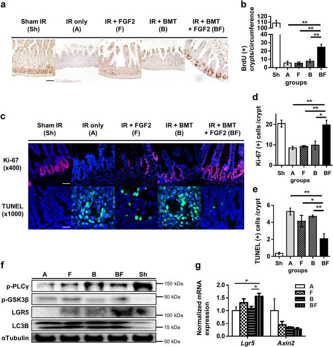
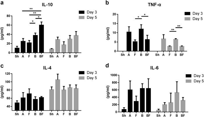
Unwanted radiological or nuclear exposure remains a public health risk for which effective therapeutic countermeasures are lacking. Here, we evaluated the efficacy of fibroblast growth factor-2 (FGF2) in treating radiation-induced gastrointestinal syndrome (RIGS) incurred by lethal whole-body irradiation (WBI) when administered in conjunction with bone marrow transplantation (BMT). In vitro experiments indicated FGF2 treatment increased proliferation, reduced apoptosis, and upregulated AKT-GSK3β/β-catenin signaling in irradiated IEC-6 cells. We next established and analyzed mice cohorts consisting of sham irradiation (Group Sh); 12 Gy WBI (Group A); WBI with BMT (Group B); WBI with FGF2 treatment (Group F); and WBI with BMT and FGF2 treatment (Group BF). At 2 weeks post-irradiation, Group BF showed a dramatic increase in survival over all other groups. Intestinal epithelium of Group BF, but not Group B or F, showed augmented proliferation, decreased apoptosis, and preserved crypt numbers and morphology. Furthermore, Group BF maintained intestinal barrier function with minimal inflammatory disturbances in a manner comparable to Group Sh. In accordance, transcriptomic analyses showed significant upregulation of intestinal barrier and stem cell markers in Group BF relative to Groups A and B. Taken together, parenteral FGF2 synergizes with BMT to confer potent mitigation against RIGS.
Conflict of interest statement
The authors declare that they have no conflict of interest.
Figures


Similar articles
-
Pretreatment with ascorbic acid prevents lethal gastrointestinal syndrome in mice receiving a massive amount of radiation.J Radiat Res. 2010;51(2):145-56. doi: 10.1269/jrr.09078. Epub 2009 Dec 4. J Radiat Res. 2010. PMID: 19959877
-
Human ghrelin mitigates intestinal injury and mortality after whole body irradiation in rats.PLoS One. 2015 Feb 11;10(2):e0118213. doi: 10.1371/journal.pone.0118213. eCollection 2015. PLoS One. 2015. PMID: 25671547 Free PMC article.
-
Bone marrow transplantation rescues intestinal mucosa after whole body radiation via paracrine mechanisms.Radiother Oncol. 2012 Dec;105(3):371-7. doi: 10.1016/j.radonc.2012.10.005. Epub 2012 Nov 9. Radiother Oncol. 2012. PMID: 23146318
-
Attenuation of radiation-induced gastrointestinal damage by epidermal growth factor and bone marrow transplantation in mice.Int J Radiat Biol. 2015;91(9):703-14. doi: 10.3109/09553002.2015.1054528. Epub 2015 Jul 3. Int J Radiat Biol. 2015. PMID: 25994811
-
Post treatment with an FGF chimeric growth factor enhances epithelial cell proliferation to improve recovery from radiation-induced intestinal damage.Int J Radiat Oncol Biol Phys. 2010 Nov 1;78(3):860-7. doi: 10.1016/j.ijrobp.2010.04.045. Epub 2010 Aug 21. Int J Radiat Oncol Biol Phys. 2010. PMID: 20729008
Cited by
-
Effects of Chinese herbal diet on hematopoiesis, immunity, and intestines of mice exposed to different doses of radiation.Heliyon. 2023 Apr 18;9(5):e15473. doi: 10.1016/j.heliyon.2023.e15473. eCollection 2023 May. Heliyon. 2023. PMID: 37131450 Free PMC article.
-
Evaluation of fibroblast growth factor-2 on the proliferation of osteogenic potential and protein expression of stem cell spheroids composed of stem cells derived from bone marrow.Exp Ther Med. 2019 Jul;18(1):326-331. doi: 10.3892/etm.2019.7543. Epub 2019 May 3. Exp Ther Med. 2019. PMID: 31258669 Free PMC article.
-
Gut microbiota-derived indole 3-propionic acid protects against radiation toxicity via retaining acyl-CoA-binding protein.Microbiome. 2020 May 20;8(1):69. doi: 10.1186/s40168-020-00845-6. Microbiome. 2020. PMID: 32434586 Free PMC article.
-
The protective effects of Xuebijing injection on intestinal injuries of mice exposed to irradiation.Animal Model Exp Med. 2022 Dec;5(6):565-574. doi: 10.1002/ame2.12285. Epub 2022 Nov 14. Animal Model Exp Med. 2022. PMID: 36376997 Free PMC article.
-
Systemic and Local Phenotypes of Barium Chloride Induced Skeletal Muscle Injury in Mice.Ann Geriatr Med Res. 2019 Jun;23(2):83-89. doi: 10.4235/agmr.19.0012. Epub 2019 Jun 30. Ann Geriatr Med Res. 2019. PMID: 32743293 Free PMC article.
References
Publication types
MeSH terms
Substances
LinkOut - more resources
Full Text Sources
Other Literature Sources
Medical
Miscellaneous
