. 2018 Mar 7;8(1):4126.
doi: 10.1038/s41598-018-22416-4.

Genomics-Based Identification of Microorganisms in Human Ocular Body Fluid

Affiliations

Genomics-Based Identification of Microorganisms in Human Ocular Body Fluid

Philipp Kirstahler et al. Sci Rep. .

Abstract

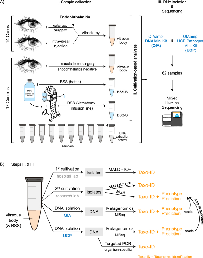
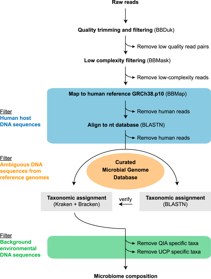
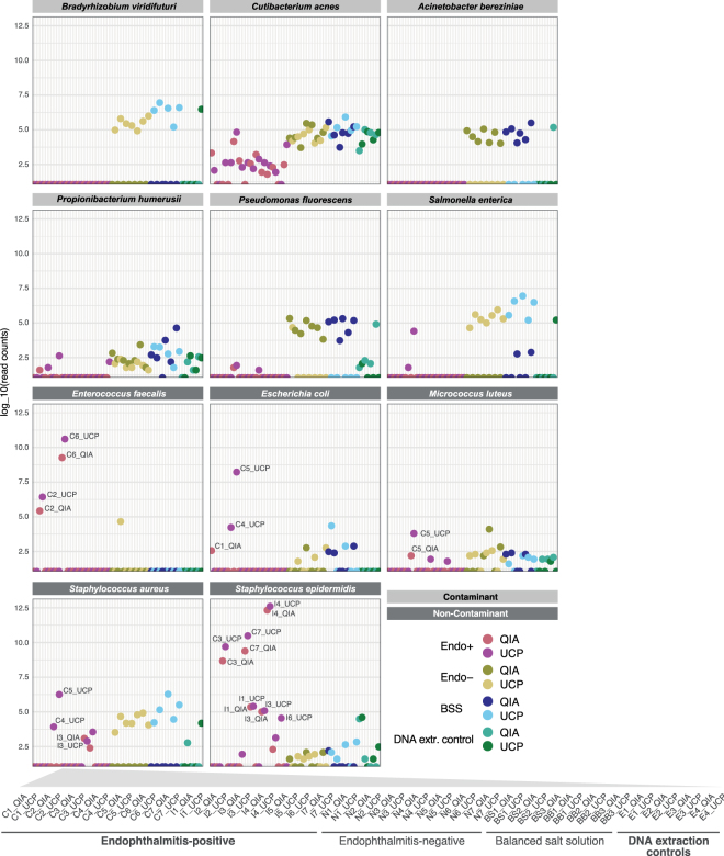
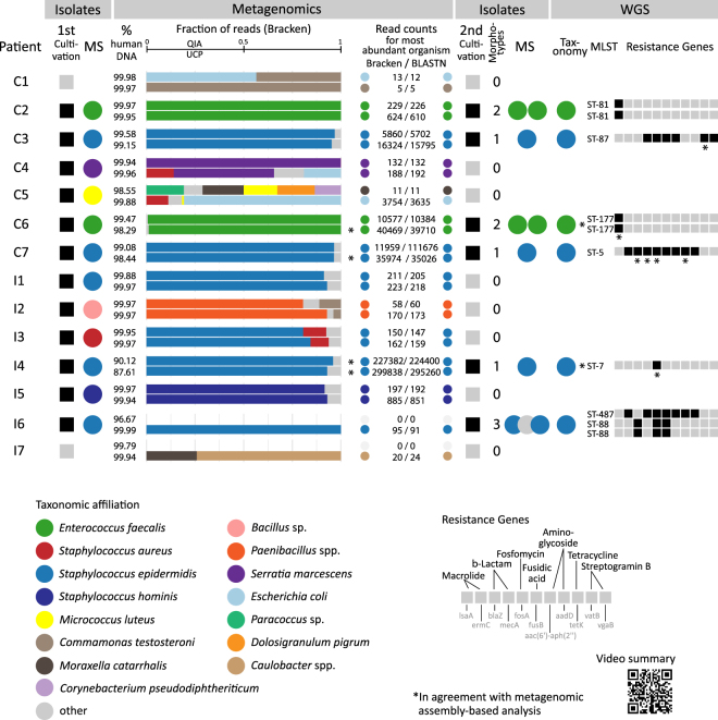
Advances in genomics have the potential to revolutionize clinical diagnostics. Here, we examine the microbiome of vitreous (intraocular body fluid) from patients who developed endophthalmitis following cataract surgery or intravitreal injection. Endophthalmitis is an inflammation of the intraocular cavity and can lead to a permanent loss of vision. As controls, we included vitreous from endophthalmitis-negative patients, balanced salt solution used during vitrectomy and DNA extraction blanks. We compared two DNA isolation procedures and found that an ultraclean production of reagents appeared to reduce background DNA in these low microbial biomass samples. We created a curated microbial genome database (>5700 genomes) and designed a metagenomics workflow with filtering steps to reduce DNA sequences originating from: (i) human hosts, (ii) ambiguousness/contaminants in public microbial reference genomes and (iii) the environment. Our metagenomic read classification revealed in nearly all cases the same microorganism that was determined in cultivation- and mass spectrometry-based analyses. For some patients, we identified the sequence type of the microorganism and antibiotic resistance genes through analyses of whole genome sequence (WGS) assemblies of isolates and metagenomic assemblies. Together, we conclude that genomics-based analyses of human ocular body fluid specimens can provide actionable information relevant to infectious disease management.


Conflict of interest statement

The authors declare no competing interests.

Figures

Figure 1
Figure 1
Sample collection, DNA isolation and shotgun metagenomic sequencing. (A) (I.) Sample collection: Vitreous body (intraocular body fluid) was collected through vitrectomy from 14 patients with endophthalmitis following cataract surgery (n = 7) and intravitreal injection (n = 7). As control, vitreous was collected from 7 patients without postoperative endophthalmitis during macula hole surgery. Six aliquots (3 sample pairs) were obtained from balanced salt solution (BSS) that is infused into the eye during vitrectomy. Three aliquots were collected from separate BSS bottles (BSS-B) and the second set of aliquots was collected from the vitrectomy surgical system (BSS-S) after it had passed through the vitrectomy infusion line, respectively. The samples were examined using (II.) Cultivation-based analyses and (III.) DNA isolation (2 methods) & Metagenomic shotgun sequencing, including the examination of DNA extraction (blank) controls. A total of 62 samples were sequenced using Illumina MiSeq sequencing technology. (B) More details to steps (II.) and (III.): (II.) Cultivation-based analyses: Aliquots of the vitreous body fluid and balanced salt solution samples were subjected to cultivation-based analyses separately at the hospital and research laboratories. Obtained isolates were analyzed using mass spectrometry and whole genome sequencing. (III.) DNA isolation & Metagenomic shotgun sequencing: Samples were extracted using two DNA isolation procedures: QIAamp DNA Mini Kit (QIA) and QIAamp UCP Pathogen Mini kit (UCP). A DNA extraction (blank) control was included at each round of DNA isolation, i.e. one DNA extraction control for 12–14 samples in total per extraction round (more vitreous samples were extracted than analyzed in this study). To verify the presence of the main microorganisms detected in the metagenomics analysis, the shotgun metagenomics reads were mapped to the genome assemblies of the isolates obtained from the vitreous samples. Not displayed here is the mapping of metagenomic shotgun reads to microbial reference genomes in the database (Provided in Fig. 4). As an additional verification, PCR analyses were carried out to detect the presence of the most abundant microorganisms in the vitreous samples using organism-specific primer sets.
Figure 2
Figure 2
Workflow for metagenomic data analysis. In a first step, sequencing adapters, low quality bases and reads with low complexity were removed. Subsequently, reads that mapped against the human reference genome sequence, or aligned with human sequences in the nt database were removed. The taxonomic classification of the reads was performed with Kraken together with Bracken using a curated microbial genome database containing 5,750 microbial (archaea [251], bacteria [5,166], fungi [225], protozoa [73], viruses [35]) and 1 human reference genome sequence (for details, see Supplementary Methods). Additional reads that in this step were classified as human were removed. To verify the classification results, the reads were also aligned to the reference genomes using BLASTn. Organisms specific for the DNA extraction (blank) controls were filtered from the patient samples.
Figure 3
Figure 3
Selected contaminant and non-contaminant organisms based on evaluation of DNA extraction control samples. Contaminant organisms (light grey) were present in higher abundance in DNA extraction controls (green) compared to the endophthalmitis-positive samples (red). The contaminant organisms were detected in similar abundance in the endophthalmitis-negative (yellow) and/or balanced salt solution samples (blue) as in the DNA extraction control samples. Organisms that were detected in higher abundance in patient samples (dark grey), compared to their respective DNA extraction control samples, were not regarded as sample contaminants. Read counts are presented as counts per million in relation to the total non-human read counts per sample, respectively. An interactive version of this figure that includes individual sample information, including read counts, is available from https://figshare.com/s/a4fd9d84260e8456ab72. For a detailed list of contaminant organisms, see Supplementary Table S5.
Figure 4
Figure 4
Coverage of bacterial reference genomes by metagenomic reads originating from intraocular fluid of endophthalmitis patients. For each individual patient, the metagenomic shotgun reads of the most abundant microbial organism were extracted at genus-level and mapped as unpaired reads using BBmap suite to the respective reference genome sequence in the database. For patients C1 and I7 a particular microbial organism could not be assigned confidently in the metagenomics analysis and these patients are regarded as “sterile” endophthalmitis cases. The outer most circle displays all sequences of the reference genome (including short contigs and plasmids). The orange and blue inner circles display the depth of mapped reads originating from the vitreous specimens that were extracted with the QIA and UCP DNA extraction methods, respectively. In the two cases where metagenomics analysis and culture results from the hospital were not identical regarding the most abundant organism (patients C5 and I3), we examined the reads via genome mapping for all organisms detected in the metagenomics analysis. The most relevant abundant organism is shown here and the additional plots, as well as information about the maximum read depth for all detected organisms, is available from https://figshare.com/s/c2ce2d32daf25db54904.
Figure 5
Figure 5
Summary of cultivation-based, metagenomics and whole genome sequence analyses. Bacterial isolates were obtained at the hospital laboratory (1st cultivation) from vitreous from endophthalmitis patients following cataract surgery (C1-7) and intravitreal injection (I1-7) and the taxonomic affiliation of the isolates were determined by MALDI-TOF mass spectrometry (MS). Vitreous was analyzed through metagenomics at the research laboratory using two DNA isolation methods (QIAamp DNA Mini Kit, QIA; QIAamp UCP Pathogen Mini kit, UCP) and the taxonomic affiliation of reads was determined. The detected amount of human DNA sequences in percent (%) is provided in the first column of the Metagenomics tab. In the horizontal bar charts, the taxonomic identity and relative fraction of microbial reads for the most abundant identified organisms based on the Kraken + Bracken analysis is indicated for both DNA isolation methods. The read counts for the most abundant organism according to the Kraken + Bracken (all reads) and BLASTN (forward read) analyses are indicated to the right. The read counts for the most abundant organisms per sample as determined by Kraken, Bracken and BLASTn analyses are available through figshare at https://figshare.com/s/5feabfad1d8c495bf7a3. Bacterial isolates for some samples were obtained in a second round of cultivation at the research laboratory (2nd cultivation) and one representative per colony morphotype per vitreous sample was subjected to MS and whole genome sequencing (WGS). The taxonomic affiliation of isolates was determined through classification of assembled genomes using a k-mer based approach and genomic MLST, and antibiotic resistance genes were identified using ResFinder. Furthermore, metagenomic assemblies were generated from the shotgun metagenomic reads and analyzed with regards to taxonomic affiliation and selected functional characteristics (Supplementary Table S6). A video summary is available from figshare at https://figshare.com/s/38fe043f6a8ef1710444.

References

    1. Durand ML. Bacterial and Fungal Endophthalmitis. Clinical Microbiology Reviews. 2017;30:597–613. doi: 10.1128/CMR.00113-16. - DOI - PMC - PubMed
    1. Bannerman TL, Rhoden DL, McAllister SK, Miller JM, Wilson LA. The source of coagulase-negative staphylococci in the Endophthalmitis Vitrectomy Study. A comparison of eyelid and intraocular isolates using pulsed-field gel electrophoresis. Arch Ophthalmol. 1997;115:357–361. doi: 10.1001/archopht.1997.01100150359008. - DOI - PubMed
    1. Buchta V, et al. Outbreak of Fungal Endophthalmitis Due to Fusarium oxysporum Following Cataract Surgery. Mycopathologia. 2014;177:115–121. doi: 10.1007/s11046-013-9721-5. - DOI - PubMed
    1. Foster A. Cataract and ‘Vision 2020—the right to sight’ initiative. British Journal of Ophthalmology. 2001;85:635–639. doi: 10.1136/bjo.85.6.635. - DOI - PMC - PubMed
    1. Solborg Bjerrum S, Kiilgaard JF, Mikkelsen KL, la Cour M. Outsourced cataract surgery and postoperative endophthalmitis. Acta Ophthalmol. 2013;91:701–708. doi: 10.1111/aos.12279. - DOI - PubMed

Publication types