Review
. 2018 Feb 20:12:84.
doi: 10.3389/fnins.2018.00084. eCollection 2018.

Steroid Transport, Local Synthesis, and Signaling within the Brain: Roles in Neurogenesis, Neuroprotection, and Sexual Behaviors

Affiliations
Review

Steroid Transport, Local Synthesis, and Signaling within the Brain: Roles in Neurogenesis, Neuroprotection, and Sexual Behaviors

Nicolas Diotel et al. Front Neurosci. .

Abstract

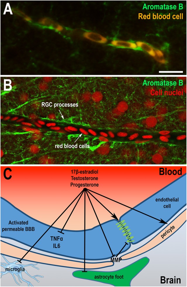
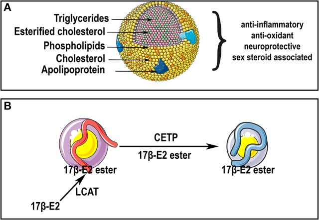
Sex steroid hormones are synthesized from cholesterol and exert pleiotropic effects notably in the central nervous system. Pioneering studies from Baulieu and colleagues have suggested that steroids are also locally-synthesized in the brain. Such steroids, called neurosteroids, can rapidly modulate neuronal excitability and functions, brain plasticity, and behavior. Accumulating data obtained on a wide variety of species demonstrate that neurosteroidogenesis is an evolutionary conserved feature across fish, birds, and mammals. In this review, we will first document neurosteroidogenesis and steroid signaling for estrogens, progestagens, and androgens in the brain of teleost fish, birds, and mammals. We will next consider the effects of sex steroids in homeostatic and regenerative neurogenesis, in neuroprotection, and in sexual behaviors. In a last part, we will discuss the transport of steroids and lipoproteins from the periphery within the brain (and vice-versa) and document their effects on the blood-brain barrier (BBB) permeability and on neuroprotection. We will emphasize the potential interaction between lipoproteins and sex steroids, addressing the beneficial effects of steroids and lipoproteins, particularly HDL-cholesterol, against the breakdown of the BBB reported to occur during brain ischemic stroke. We will consequently highlight the potential anti-inflammatory, anti-oxidant, and neuroprotective properties of sex steroid and lipoproteins, these latest improving cholesterol and steroid ester transport within the brain after insults.

Keywords: HDL; aromatase; blood-brain barrier; cholesterol; estrogens; lipoproteins; progestins; stroke.


Figures

Figure 1
Figure 1
Biosynthetic pathways of neurosteroid synthesis. Cyp11a1, cytochrome P450 family 11 subfamily A member 1 (P450scc), cytochrome P450 side-chain cleavage; Cyp17, cytochrome P450 17α-hydroxylase/C17, 20-lyase; Aro, aromatase; 3β-HSD, 3β-hydroxysteroid dehydrogenase D5–D4 isomerase; 5α-R, 5α-reductase; 17β-HSD, 17β-hydroxysteroid dehydrogenase; Cyp21, cytochrome P450 21 hydroxylase; Cyp11, cytochrome P450 11β-hydroxylase B1 and/or B2. StAR, steroidogenic acute regulatory protein.
Figure 2
Figure 2
Steroidogenesis in the brain of fish and mammals. (A) Steroidogenesis in the brain of adult fish. The blue cell corresponds to a radial glia cell acting as neural stem cell, while the green cell corresponds to a neuron. Data obtained by in situ hybridization, RNA sequencing and/or proteomics show neuronal and radial glial expression of steroidogenic enzymes involved in the synthesis of 17OH-Pregnenolone (17OH-P), Progesterone (P), dehydroepiandrosterone (DHEA), Androstenedione (Andro), Estrone (E1), 17β-estradiol (17β-E2, called here E2), testosterone (T) and dihydro-testosterone (DHT), cortisol and THDOC (tetrahydrodeoxycorticosterone). (B) Steroidogenesis in the brain of rat. The purple cell corresponds to an oligodendrocyte that participates in the myelin sheath genesis of axons; the green cell is an astrocyte and the yellow one is a neuron whose axon is myelinated by the oligodendrocyte. This schematic view adapted from Zwain and Yen (1999) highlights potential interaction of oligodendrocytes, astrocytes, and neurons in neurosteroidogenesis. The main steroidogenic pathway is shown by black arrows, the minor ones by red arrows and the suggested ones by blue arrows referring to the work of Zwain and Yen (1999). Note that arrows do not necessarily document the direct conversion of the one steroid into another, but can include several enzymatic processes.
Figure 3
Figure 3
Potential roles of steroids in the brain. (A) Effects of 17β-estradiol on neurogenesis in adult zebrafish. At the top, the scheme represents a zebrafish and a transversal brain section through the olfactory bulbs, taken from Menuet et al. (2005). The lines on the right hemisphere correspond to radial glial cells, that behave as neural stem cells. At the bottom, the scheme illustrates neurogenesis events occurring on the zebrafish brain. Radial glial cells (blue) give rise to new neurons (green) that migrate and differentiate into mature and functional neurons (yellow). The orange cells close to the ventricle vicinity are further committed progenitors. It was shown that 17β-estradiol impairs neural progenitor proliferation and new born cell migration; the data obtained for neural differentiation and survival are not clear (Diotel et al., ; Pellegrini et al., 2015). (B) Effects of sex steroids on neurogenesis in adult rodents. At the top, the scheme represents a rodent and a transversal brain section through the dentate gyrus (DG) of the hippocampus (framed box), where neurogenesis is maintained during adulthood. At the bottom, the scheme illustrates neurogenesis events occurring in the granular cell layer of the dentate gyrus. Radial glial cells (blue light) give rise to neural progenitors (orange) that give birth to new neurons (green with a small neuritic arborescence) that migrate and differentiate into mature and functional neurons (green with a huge neuritic arborescence). In general, 17β-estradiol (17β-E2) favors neural stem cell/progenitor proliferation; to a lesser extent, testosterone (T) and progesterone (P) also exert positive role on proliferation; 17β-estradiol, testosterone and progesterone also promote neuronal migration and differentiation (Heberden, 2017). In addition, these three sex steroids promote new born cell survival in constitutive and regenerative neurogenesis. Note, that the effects of sex steroids on neurogenesis could be dependent of the timing, the concentration, the rhythm of exposure and the regions targeted. For instance, estradiol treatment has been shown to decrease neurogenesis in the SVZ (Brock et al., 2010). (C) Peripheral steroids and neurosteroids impact brain functions and homeostasis, by modulating neurogenesis under homeostatic and regenerative conditions, by promoting neuroprotection, learning, and memory, and by exerting anti-inflammatory and antioxidant properties. They also modulate sexual behavior.
Figure 4
Figure 4
Sex hormones restore blood-brain barrier physiology and integrity. (A,B) Aromatase B immunohistochemistry (green) on zebrafish brain highlights that radial glial processes envelop blood vessels as shown by the auto-fluorescence of red blood cells (A) or by nuclear staining (B). These data suggest that radial glial cells could endorse the role of astrocytes in the establishment of the BBB, given the absence of astrocytic cell-type in the brain of fish. It also raises the question of the potential role of locally-produced estrogens on the BBB physiology. Scale bar: 7 μm. (C) Sex steroids (17β-E2, progesterone, and testosterone) display direct and indirect effects on the BBB through the restauration of thigh junctions, the inhibition of inflammatory cytokine expression and metalloproteinases production, the regulation of pericytes contraction, and consequently the modulation of cerebral blood flow. They also limit reactive gliosis through the inhibition of glial activation under pathological conditions (astrocytes and microglia). Sex steroids participate in the maintenance of a functional BBB, reducing neuroinflammation and promoting neuroprotection.
Figure 5
Figure 5
HDL particles and sex steroids. (A) Lipoprotein particles display a central core containing cholesterol, cholesterol esters and triglycerides, that is enveloped by free cholesterol, phospholipids, and apolipoproteins. HDL particles display pleiotropic effects partly supported by their anti-inflammatory, anti-oxidant and neuroprotective properties. HDLs have been shown to display metabolic interaction with sex steroids (i.e., androstenediol, estradiol, DHEA, dihydrotestosterone, pregnenolone, and progesterone). (B) As suggested by Tikkanen et al. (2002), 17β-E2 could be esterified with fatty acids by lecithin:cholesterol acyltransferase (LCAT) in HDL. Esterified 17β-E2 could be subsequently transferred from HDLs to LDLs by cholesteryl ester transfer protein (CETP). Thus, HDL particles could be a source of cholesterol and steroids in the brain notably after CNS insults, and could be used as a carrier for improving steroids delivery in therapeutically approaches.

References

    1. Adolf B., Chapouton P., Lam C. S., Topp S., Tannhäuser B., Strähle U., et al. . (2006). Conserved and acquired features of adult neurogenesis in the zebrafish telencephalon. Dev. Biol. 295, 278–293. 10.1016/j.ydbio.2006.03.023 - DOI - PubMed
    1. Alafuzoff I., Adolfsson R., Bucht G., Winblad B. (1983). Albumin and immunoglobulin in plasma and cerebrospinal fluid, and blood-cerebrospinal fluid barrier function in patients with dementia of Alzheimer type and multi-infarct dementia. J. Neurol. Sci. 60, 465–472. 10.1016/0022-510X(83)90157-0 - DOI - PubMed
    1. Alkayed N. J., Harukuni I., Kimes A. S., London E. D., Traystman R. J., Hurn P. D. (1998). Gender-linked brain injury in experimental stroke. Stroke 29, 159–165: discussion 166. - PubMed
    1. Alunni A., Vaccari S., Torcia S., Meomartini M. E., Nicotra A., Alfei L. (2005). Characterization of glial fibrillary acidic protein and astroglial architecture in the brain of a continuously growing fish, the rainbow trout. Eur. J. Histochem. 49, 157–166. - PubMed
    1. Andersson E., Borg B., Lambert J. G. (1988). Aromatase activity in brain and pituitary of immature and mature Atlantic salmon (Salmo salar L.) parr. Gen. Comp. Endocrinol. 72, 394–401. 10.1016/0016-6480(88)90161-X - DOI - PubMed

LinkOut - more resources