. 2018 Feb 20:11:45.
doi: 10.3389/fnmol.2018.00045. eCollection 2018.

CB1R-Mediated Activation of Caspase-3 Causes Epigenetic and Neurobehavioral Abnormalities in Postnatal Ethanol-Exposed Mice

Affiliations

CB1R-Mediated Activation of Caspase-3 Causes Epigenetic and Neurobehavioral Abnormalities in Postnatal Ethanol-Exposed Mice

Shivakumar Subbanna et al. Front Mol Neurosci. .

Abstract

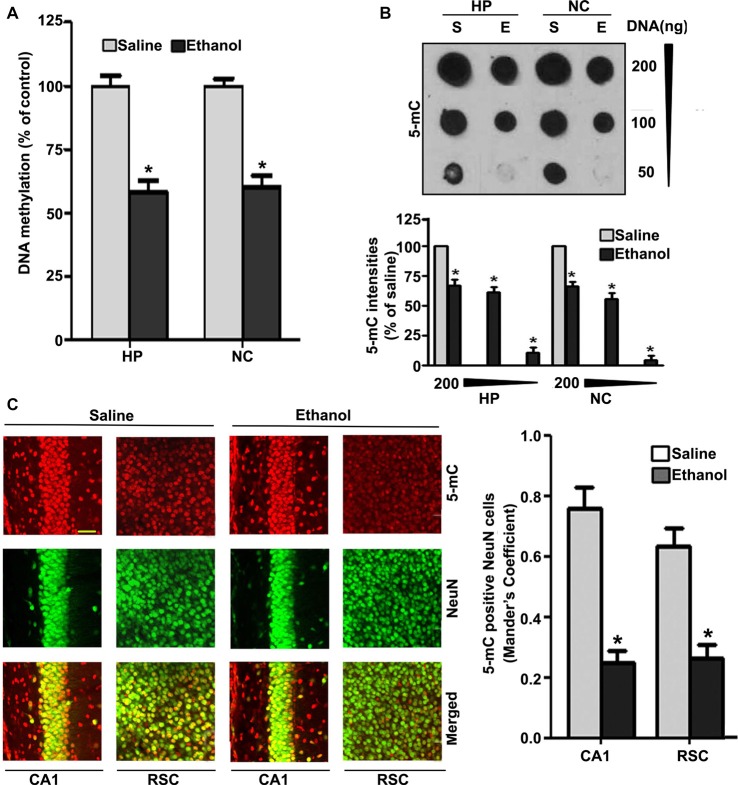
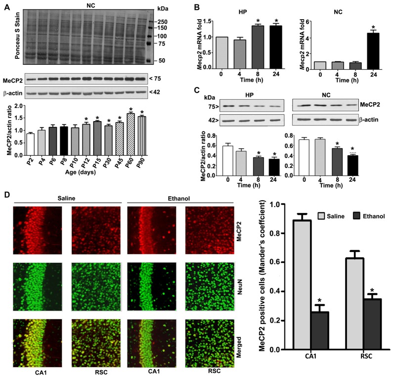
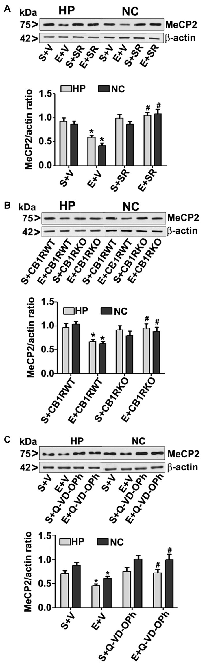
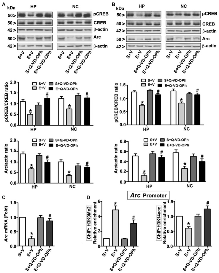
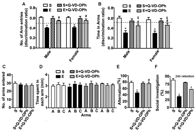
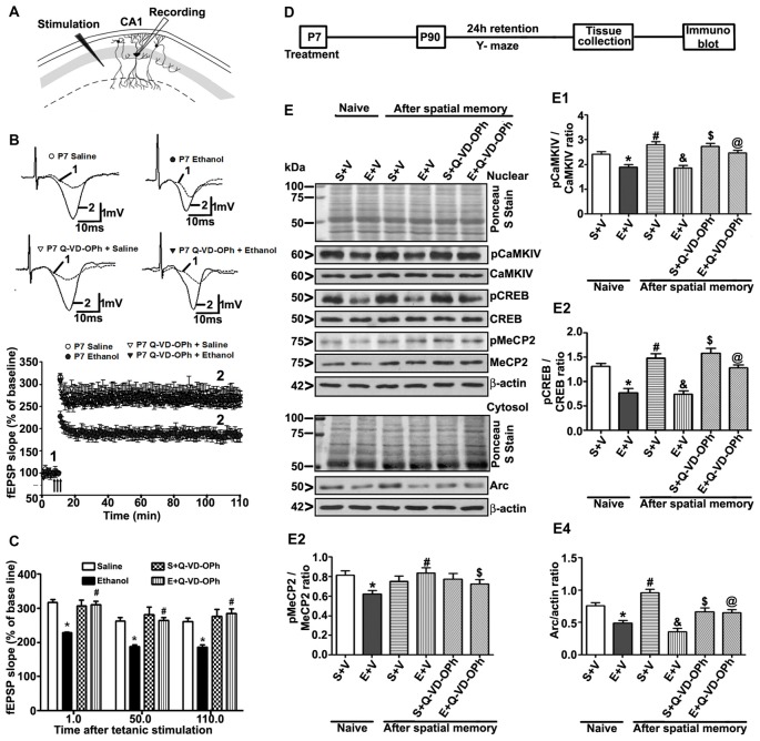
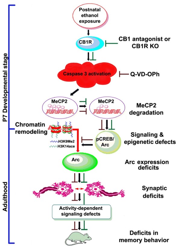
Alcohol exposure can affect brain development, leading to long-lasting behavioral problems, including cognitive impairment, which together is defined as fetal alcohol spectrum disorder (FASD). However, the fundamental mechanisms through which this occurs are largely unknown. In this study, we report that the exposure of postnatal day 7 (P7) mice to ethanol activates caspase-3 via cannabinoid receptor type-1 (CB1R) in neonatal mice and causes a reduction in methylated DNA binding protein (MeCP2) levels. The developmental expression of MeCP2 in mice is closely correlated with synaptogenesis and neuronal maturation. It was shown that ethanol treatment of P7 mice enhanced Mecp2 mRNA levels but reduced protein levels. The genetic deletion of CB1R prevented, and administration of a CB1R antagonist before ethanol treatment of P7 mice inhibited caspase-3 activation. Additionally, it reversed the loss of MeCP2 protein, cAMP response element binding protein (CREB) activation, and activity-regulated cytoskeleton-associated protein (Arc) expression. The inhibition of caspase-3 activity prior to ethanol administration prevented ethanol-induced loss of MeCP2, CREB activation, epigenetic regulation of Arc expression, long-term potentiation (LTP), spatial memory deficits and activity-dependent impairment of several signaling molecules, including MeCP2, in adult mice. Collectively, these results reveal that the ethanol-induced CB1R-mediated activation of caspase-3 degrades the MeCP2 protein in the P7 mouse brain and causes long-lasting neurobehavioral deficits in adult mice. This CB1R-mediated instability of MeCP2 during active synaptic maturation may disrupt synaptic circuit maturation and lead to neurobehavioral abnormalities, as observed in this animal model of FASD.

Keywords: DNA methylation; FASD; MeCP2; neurodegeneration; synaptic plasticity.


Figures

Figure 1
Figure 1
Treatment of postnatal day 7 (P7) mice with ethanol reduces DNA methylation in the brain. (A) Global DNA methylation was reduced by ethanol treatment. Global DNA methylation quantification in DNA from the hippocampus (HP) and neocortex (NC) obtained 8 h after the first saline or ethanol injection. For the 0 h ethanol group, saline was injected. (*p < 0.05, n = 10 pups/group). (B) Global 5-mC-specific dot-blot intensities of genomic DNA prepared from the HP and NC (200 ng, 100 ng and 50 ng of genomic DNA) of P7 mice after treatment with saline or ethanol (8 h). The 5-mC-specific dot-blot intensities (%) compared with the saline controls (*p < 0.01 vs. Saline, n = 8 pups/group). (C) Free-floating coronal brain sections (HP and RSC) from both the groups (saline and 8 h ethanol) were subjected to immunohistochemistry with anti-mouse 5mC and anti-rabbit NeuN antibodies to label 5-mC-positive NeuN in neurons. Mander’s coefficient analysis was used to evaluate 5-mC-positive NeuN neurons in the CA1 and RSC brain regions. (*p < 0.01 vs. Saline, n = 6 pups/group). Scale bars = 10 μm. Error bars, SEM (one-way analysis of variance (ANOVA) with Bonferroni’s post hoc test).
Figure 2
Figure 2
MeCP2 protein expression pattern during mouse brain development; ethanol treatment of P7 mice enhances Mecp2 mRNA but impairs the protein levels in the HP and NC regions. (A) MeCP2 protein expression pattern during mouse brain development. The MeCP2 expression pattern in neocortical nuclear protein extract obtained from P2 to P90 mouse brains were subjected to western blotting. Equal protein loading was confirmed after Ponceau S staining, and β-actin was used as the protein loading control. (*p < 0.01 vs P2 groups; n = 12 pups/group). (B) RT-qPCR analysis of Mecp2 mRNA in HP and NC extracts obtained 4–24 h after the first saline or ethanol injection (n = 12 pups/group). Gapdh mRNA was used as the internal control for normalization of Mecp2 mRNA levels. (C) Western blot analysis of MeCP2 proteins in the P7 HP and NC nuclear protein extracts (4–24 h after the first saline or ethanol treatment). β-actin was used as the protein loading control. In the 0 h ethanol group, saline was injected [*p < 0.01 vs. saline (0 h) group]. (n = 12 pups/group). (D) The coronal brain sections (HP, hippocampus, and RSC, retrosplenial cortex; saline and 8 h ethanol) were used for immunohistochemistry with anti-mouse MeCP2 and anti-rabbit NeuN antibodies to label MeCP2-positive NeuN in neurons. Mander’s coefficient analysis was used to evaluate MeCP2-positive NeuN neurons in the CA1 and RSC brain regions. Error bars, SEM (*p < 0.01 vs. Saline, n = 6 pups/group). Scale bars = 10 μm. Error bars, SEM (one-way ANOVA with Bonferroni’s post hoc test).
Figure 3
Figure 3
CB1R antagonist (SR141716A) or CB1R knockout (KO) mice or Q-VD-OPh protect against ethanol-mediated loss of MeCP2 proteins. (A) P7 mice were pretreated with SR (1 mg/kg) 30 min before saline or ethanol treatment. MeCP2 proteins from hippocampal (HP) and neocortical (NC) nuclear extracts from the four treatment groups (S + V, E + V, S + SR and E + SR) groups (n = 12 pups/group) were subjected to western blotting. S, saline; E, ethanol. β-actin was used as the protein loading control. (*p < 0.01 vs. S + V; #p < 0.01 vs. E + V). (B) P7 CB1R wild-type (WT) and CB1R KO mice were treated with saline or ethanol. From nuclear extracts of HP and NC, western blot analysis of MeCP2 protein was performed for all the four treatment groups (S + CB1R WT, E + CB1R WT, S + CB1R KO + and E + CB1R KO) groups (n = 12 pups/group). S, saline; E, ethanol. (*p < 0.01 vs. S + CB1RWT; #p < 0.01 vs. E + CB1RWT). (C) P7 mice pre-treated for 30 min with Q-VD-OPh (1 mg/kg) or vehicle were exposed to ethanol for 8 h, and MeCP2 protein levels were examined in nuclear extracts of the HP and NC from saline (S), ethanol (E), S + Q-VD-OPh, or E + Q-VD-OPh groups (n = 12 pups/group) via western blot analysis. β-actin was used as the protein loading control (*p < 0.01 vs. S; #p < 0.01 vs. E. Error bars, SEM (two-way ANOVA with Bonferroni’s post hoc test).
Figure 4
Figure 4
Pharmacological blockades of caspase-3 activation protects against ethanol-induced inhibition of CREB phosphorylation and epigenetic-mediated activity regulatedcytoskeleton-associated protein (Arc) expression in the mouse brain. P7 pups were injected with Q-VD-OPh (1 mg/kg) 30 min before first saline or ethanol exposure and were either sacrificed after 8 h or were allowed to mature to adulthood and then sacrificed. (A) P7 and (B) adult hippocampal (HP) and neocortical (NC) nuclear extracts from the four treatment groups (S + V, E + V, S + Q-VD-OPh, or E + Q-VD-OPh) were subjected to western blotting to analyze the levels of pCREB, CREB and Arc (n = 10 pups/group). The representative blots are shown for the hippocampal and cortical nuclear extracts (*p < 0.01 vs. S + V; #p < 0.01 vs. E + V). β-actin was used as the loading control. (C) Adult hippocampal nuclear extracts from the four treatment groups (S + V, E + V, S + Q-VD-OPh, or E + Q-VD-OPh) were subjected to RT-qPCR to analyze the Arc mRNA levels. Hprt mRNA was used as the internal control for normalization of Arc mRNA. (D) Epigenetic analysis at the promoter region of the Arc gene. ChIP analysis of the Arc gene promoter in HP tissues from the four treatment groups (S + V, E + V, S + Q-VD-OPh, or E + Q-VD-OPh) with anti-acetylated H3K14 or anti-H3K9me2 antibodies. Levels of Arc gene promoter chromatin enrichment in the IPs were measured by RT-qPCR. Error bars, SEM (two-way ANOVA with Bonferroni’s post hoc test).
Figure 5
Figure 5
Administration of a caspase-3 inhibitor before ethanol treatment in P7 mice rescues persistent spatial and social recognition memory (SRM) deficits in adulthood. (A,B) The spatial working memory was determined using the Y-maze in adult mice treated with saline (S), ethanol (E), S + Q-VD-OPh, or E + Q-VD-OPh at P7. The discrimination ratio [preference for the novel arm over the familiar other arm (Novel/Novel + Other)] for arms entries (A) and dwell time (time spent in each arm) (B) of S, E, S + Q-VD-OPh and E + Q-VD-OPh -treated mice, 24 h after the first encounter with the partially opened maze. The dashed line denotes chance performance (0.5). (C–E) The spontaneous alternation memory of adult mice treated with saline (S), ethanol (E), S + Q-VD-OPh, or E + Q-VD-OPh at P7 was evaluated using the Y-maze. (C) The number of arm entries by mice in all the treatment groups. (D) The time spent in each arm by mice in all the treatment groups. (E) The spontaneous alternation performance by mice in all the treatment groups. (F) The percent of social investigation is shown for P7 S, E, S + Q-VD-OPh and E + Q-VD-OPh -treated adult mice, 24 h after the first encounter with the same juvenile mice. (*p < 0.01 vs. saline; $p < 0.05 vs. saline; #p < 0.05 vs. ethanol, n = 8 mice/group). Error bars, SEM (two-way ANOVA with Bonferroni’s post hoc test).
Figure 6
Figure 6
Administration of a caspase-3 inhibitor before P7 ethanol treatment prevents long-term potentiation (LTP) deficits in adult male mice. (A) A schematic drawing shows the stimulating and recording electrode positions in the CA1 region of the HP. (B) The average field-excitatory-post-synaptic potential (fEPSP) slope at various time points obtained from P7 saline (S), ethanol (E), S + Q-VD-OPh, or E + Q-VD-OPh-treated adult mice. For each slice, the fEPSP slopes were normalized against the average slope over the 10-min recording before LTP stimulation. Arrows show the time of Theta-Burst Stimulation (TBS; four pulses at 100 Hz, with bursts repeated at 5 Hz, and each tetanus including three different 10-burst trains separated by 15 s). Inset: representative traces before and after TBS are shown for all the treatment groups. Scale 1 mV; 10 ms. (C) Bar graph compares the average of the fEPSP slopes at several time points after TBS in P7 saline (S), ethanol (E), S + Q-VD-OPh, or E + Q-VD-OPh-treated adult mice hippocampal slices. (*p < 0.01 vs. saline, n = 5 mice/group; 10 slices/group). (D) Scheme indicating the experimental design to evaluate the effects of pre-administration of Q-VD-OPh before saline/ethanol treatment in P7 mice on activity-dependent signaling events in adulthood. The spatial working memory of adult mice treated with saline (S), ethanol (E), S + Q-VD-OPh, or E + Q-VD-OPh at P7 was evaluated using a Y-maze. (E) HP tissues were collected immediately after a 24 h intertrial interval when the mice completed exploring all three arms (3 min, preference trial, test trial). The signaling proteins (E1) pCaMKIV, (E2) pCREB, (E3) pMeCP2 and (E4) Arc were examined in nuclear or cytosolic extracts of the HP from saline (S), ethanol (E), S + Q-VD-OPh, or E + Q-VD-OPh groups (n = 8 mice/group) via western blot analysis. Equal protein loading was confirmed after Ponceau S staining, and β-actin was used as a protein loading control (*p < 0.01 vs. S; #p < 0.01 vs. E; &p < 0.01 vs. S+V; $p < 0.01 vs S; @p < 0.01 vs E. Error bars, SEM (two-way ANOVA with Bonferroni’s post hoc test).
Figure 7
Figure 7
Schematic diagram showing the molecular mechanism by which CB1R-mediated activation of caspase-3 degrades MeCP2, leading to neurobehavioral abnormalities in postnatal ethanol-exposed mice. It was revealed previously that P7 ethanol treatment activates CB1R and inhibits pCREB and Arc expression, leading to neurodegeneration in neonatal mice and persistent neurobehavioral abnormalities in adult mice (Subbanna et al., 2015a). Here, we show that inhibition of CB1R (Subbanna et al., 2013a, 2014) or caspase-3 prevents loss of MeCP2 and rescues pCREB/Arc defects in neonatal mice. Inhibition of caspase-3 in P7 mice also protected against the synaptic plasticity, Arc expression, activity-dependent signaling and behavioral defects in adult mice exposed to ethanol at P7. These observations suggest that CB1R (Subbanna et al., 2013a, 2014)/caspase-3-mediated MeCP2 loss in early development causes neurobehavioral abnormalities in postnatal ethanol-exposed adult mice (↓, ethanol effects; ⊥, drug effects).

Similar articles

Cited by

References

    1. Abel E. L., Jacobson S., Sherwin B. T. (1983). In utero alcohol exposure: functional and structural brain damage. Neurobehav. Toxicol. Teratol. 5, 363–366. - PubMed
    1. Akbarian S., Chen R. Z., Gribnau J., Rasmussen T. P., Fong H., Jaenisch R., et al. . (2001). Expression pattern of the Rett syndrome gene MeCP2 in primate prefrontal cortex. Neurobiol. Dis. 8, 784–791. 10.1006/nbdi.2001.0420 - DOI - PubMed
    1. Alati R., Al Mamun A., Williams G. M., O’Callaghan M., Najman J. M., Bor W. (2006). In utero alcohol exposure and prediction of alcohol disorders in early adulthood: a birth cohort study. Arch. Gen. Psychiatry 63, 1009–1016. 10.1001/archpsyc.63.9.1009 - DOI - PubMed
    1. Amir R. E., van den Veyver I. B., Wan M., Tran C. Q., Francke U., Zoghbi H. Y. (1999). Rett syndrome is caused by mutations in X-linked MECP2, encoding methyl-CpG-binding protein 2. Nat. Genet. 23, 185–188. 10.1038/13810 - DOI - PubMed
    1. Arancio O., Kandel E. R., Hawkins R. D. (1995). Activity-dependent long-term enhancement of transmitter release by presynaptic 3′,5′-cyclic GMP in cultured hippocampal neurons. Nature 376, 74–80. 10.1038/376074a0 - DOI - PubMed

LinkOut - more resources