Cell Behaviors during Closure of the Choroid Fissure in the Developing Eye
- PMID: 29515375
- PMCID: PMC5826230
- DOI: 10.3389/fncel.2018.00042
Cell Behaviors during Closure of the Choroid Fissure in the Developing Eye
Abstract
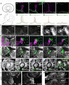
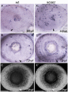
Coloboma is a defect in the morphogenesis of the eye that is a consequence of failure of choroid fissure fusion. It is among the most common congenital defects in humans and can significantly impact vision. However, very little is known about the cellular mechanisms that regulate choroid fissure closure. Using high-resolution confocal imaging of the zebrafish optic cup, we find that apico-basal polarity is re-modeled in cells lining the fissure in proximal to distal and inner to outer gradients during fusion. This process is accompanied by cell proliferation, displacement of vasculature, and contact between cells lining the choroid fissure and periocular mesenchyme (POM). To investigate the role of POM cells in closure of the fissure, we transplanted optic vesicles onto the yolk, allowing them to develop in a situation where they are depleted of POM. The choroid fissure forms normally in ectopic eyes but fusion fails in this condition, despite timely apposition of the nasal and temporal lips of the retina. This study resolves some of the cell behaviors underlying choroid fissure fusion and supports a role for POM in choroid fissure fusion.
Keywords: choroid fissure; coloboma; eye; morphogenesis; optic fissure; optic vesicle; periocular mesenchyme; zebrafish.
Figures


Similar articles
-
Retinoic acid receptor signaling regulates choroid fissure closure through independent mechanisms in the ventral optic cup and periocular mesenchyme.Proc Natl Acad Sci U S A. 2011 May 24;108(21):8698-703. doi: 10.1073/pnas.1103802108. Epub 2011 May 9. Proc Natl Acad Sci U S A. 2011. PMID: 21555593 Free PMC article.
-
The cellular bases of choroid fissure formation and closure.Dev Biol. 2018 Aug 15;440(2):137-151. doi: 10.1016/j.ydbio.2018.05.010. Epub 2018 May 24. Dev Biol. 2018. PMID: 29803644 Free PMC article.
-
Pax2a, but not pax2b, influences cell survival and periocular mesenchyme localization to facilitate zebrafish optic fissure closure.Dev Dyn. 2022 Apr;251(4):625-644. doi: 10.1002/dvdy.422. Epub 2021 Sep 28. Dev Dyn. 2022. PMID: 34535934 Free PMC article.
-
Genes and pathways in optic fissure closure.Semin Cell Dev Biol. 2019 Jul;91:55-65. doi: 10.1016/j.semcdb.2017.10.010. Epub 2017 Dec 6. Semin Cell Dev Biol. 2019. PMID: 29198497 Review.
-
Embryology of the eye and its adnexae.Dev Ophthalmol. 1992;24:1-142. Dev Ophthalmol. 1992. PMID: 1628748 Review.
Cited by
-
Eye morphogenesis in the blind Mexican cavefish.Biol Open. 2021 Oct 15;10(10):bio059031. doi: 10.1242/bio.059031. Epub 2021 Oct 28. Biol Open. 2021. PMID: 34590124 Free PMC article.
-
Closing the Gap: Mechanisms of Epithelial Fusion During Optic Fissure Closure.Front Cell Dev Biol. 2021 Jan 11;8:620774. doi: 10.3389/fcell.2020.620774. eCollection 2020. Front Cell Dev Biol. 2021. PMID: 33505973 Free PMC article. Review.
-
Genetics of congenital eye malformations: insights from chick experimental embryology.Hum Genet. 2019 Sep;138(8-9):1001-1006. doi: 10.1007/s00439-018-1900-5. Epub 2018 Jul 6. Hum Genet. 2019. PMID: 29980841 Review.
-
Nf2 fine-tunes proliferation and tissue alignment during closure of the optic fissure in the embryonic mouse eye.Hum Mol Genet. 2020 Dec 18;29(20):3373-3387. doi: 10.1093/hmg/ddaa228. Hum Mol Genet. 2020. PMID: 33075808 Free PMC article.
-
Detailed analysis of chick optic fissure closure reveals Netrin-1 as an essential mediator of epithelial fusion.Elife. 2019 Jun 4;8:e43877. doi: 10.7554/eLife.43877. Elife. 2019. PMID: 31162046 Free PMC article.
References
Grants and funding
LinkOut - more resources
Full Text Sources
Other Literature Sources
Molecular Biology Databases

