. 2018 Feb 20:9:262.
doi: 10.3389/fmicb.2018.00262. eCollection 2018.

Staphylococcus aureus Membrane-Derived Vesicles Promote Bacterial Virulence and Confer Protective Immunity in Murine Infection Models

Affiliations

Staphylococcus aureus Membrane-Derived Vesicles Promote Bacterial Virulence and Confer Protective Immunity in Murine Infection Models

Fatemeh Askarian et al. Front Microbiol. .

Abstract

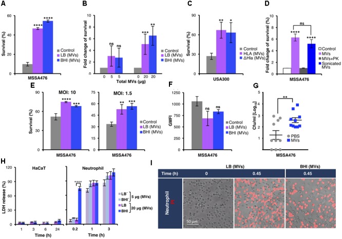
Staphylococcus aureus produces membrane-derived vesicles (MVs), which share functional properties to outer membrane vesicles. Atomic force microscopy revealed that S. aureus-derived MVs are associated with the bacterial surface or released into the surrounding environment depending on bacterial growth conditions. By using a comparative proteomic approach, a total of 131 and 617 proteins were identified in MVs isolated from S. aureus grown in Luria-Bertani and brain-heart infusion broth, respectively. Purified S. aureus MVs derived from the bacteria grown in either media induced comparable levels of cytotoxicity and neutrophil-activation. Administration of exogenous MVs increased the resistance of S. aureus to killing by whole blood or purified human neutrophils ex vivo and increased S. aureus survival in vivo. Finally, immunization of mice with S. aureus-derived MVs induced production of IgM, total IgG, IgG1, IgG2a, and IgG2b resulting in protection against subcutaneous and systemic S. aureus infection. Collectively, our results suggest S. aureus MVs can influence bacterial-host interactions during systemic infections and provide protective immunity in murine models of infection.

Keywords: Staphylococcus aureus; membrane-derived vesicles; protective immunity; proteomics; systemic infection.


Figures

FIGURE 1
FIGURE 1
The size and protein cargo of S. aureus-derived MVs were markedly different depending on growth conditions. (A) TEM-immunogold analysis of MV-generation on S. aureus MSSA476 (black arrow) grown on LA and blood agar plate. Gold particles (white arrows) indicate the presence of peptidoglycan/peptidoglycan-precursors. The scale bar is shown. (B) Atomic force micrographs of S. aureus MSSA476 cultivated on LA, BHI- and blood agar plates. Arrows indicate MVs on the bacterial surface and the released MVs. The scale bar is shown. (C) The average size distribution of MVs was determined using dynamic light scattering. The data are expressed as mean ± SEM (standard error of the mean) of four independent measurements performed on independent samples. (D) TEM-immunogold labeling of peptidoglycan/peptidoglycan-precursors in purified vesicles (black arrows) obtained from S. aureus MSSA476 grown in BHI and LB purified by flotation through an OptiPrep density gradient. Gold particles (white arrows) indicate the presence of peptidoglycan/peptidoglycan-precursors. Scale bars are shown. (E) S. aureus-derived vesicles from MSSA476 grown in LB and BHI were purified by flotation through an OptiPrep density gradient. Venn diagram illustrates common and unique proteins associated with MV identified using LPI/in-solution approaches. (F) Localization of the MV-associated proteins shown as the percentage in a pie chart. (G) Histograms representing the distribution of identified vesicular proteins according to their molecular functions, role in biological processes and cellular components. (H) Supplementation of MVs (20 μg of total MV, i.e., 0.1 μg/μl) in the culture media did not influence bacterial growth in LB media (left panel) or BHI (right panel). Data represent as the mean ± SEM of three independent experiments. The significance is indicated by asterisks (): ∗∗P ≤ 0.01.
FIGURE 2
FIGURE 2
Staphylococcus aureus MVs promote bacterial survival in human whole blood and in the presence of neutrophils ex vivo and in vivo. (A) Survival of S. aureus MSSA476 in blood is increased in the presence of 20 μg MV (0.1 μg/μl) isolated from MSSA476 grown in LB and BHI [marked as LB (MVs) or BHI (MVs) in the figure] compared to absence of MVs (marked as control in the figure). The number of inoculated bacteria at time point 0 was set to 100% and the number of surviving bacteria after 3 h is represented as the percentage of inoculation. (B) S. aureus MSSA476 survival in blood is increased in the presence of MVs, in a dose-dependent manner (5–20 μg of total MVs, i.e., 0.025–0.1 μg/μl). The number of surviving bacteria after 3 h in the absence of MVs was arbitrary set as 1, and the number of surviving bacteria in the presence of MVs is represented as the fold change compared to bacteria in the absence of MV. (C) Survival of USA300 MRSA in human blood is increased in the presence of MVs isolated from USA300 (MV-Hla) and USA300ΔHla (MV-ΔHla) grown in BHI. The percentage of survival was calculated as described in (A). (D) Sonication of purified MVs followed by proteinase K (PK) treatment abolished the effect of MVs on bacterial survival in human whole blood. The fold change of survival was calculated as described in (B). (E) Survival of opsonized S. aureus MSSA476 in the presence of neutrophils is enhanced by supplementation of MVs isolated from bacteria growing in LB and BHI. Percentage of survival was calculated as described in (A). (F) S. aureus MSSA476 were labeled with FITC and incubated with human whole blood in the absence or presence of MVs isolated from MSSA476 grown in LB and BHI. Data represents geometric mean of the fluorescence intensity (GMFI). (G) Bacterial loads in the blood (CFU/ml) of 8-week-old C57BL/6 mice were counted 24 h after the mice were intravenously infected with S. aureus MSSA476 supplemented with PBS or an exogenous source of MVs isolated from MSSA476 grown in BHI. (H) HaCaT (100 μg of total MVs, i.e., 0.1 μg/μl) and freshly purified neutrophils were treated with MVs (5–20 μg of total MVs, i.e., 0.025–0.1 μg/μl) isolated from S. aureus MSSA476 grown in LB or BHI at the time points indicated. Percentage of cytotoxicity was calculated by measuring the amount of LDH released from the cytosol of damaged cells into the supernatant after exposure to MVs. (I) Viability staining of neutrophils in the presence (20 μg of total MVs, i.e., 0.1 μg/μl) or absence of MVs were performed using propidium iodide (PI). Live imaging was performed after 0 and 0.45 or 1.5 h using fluorescence microscopy. Scale bar is shown. The data represent as the mean ± SEM of at least three independent experiments except for (D), which the data are expressed as the mean ± SEM of two independent experiments performed in triplicate. Mice study corresponds to one experiment performed with 10 mice/group. The significance is indicated by asterisks (): P < 0.05; ∗∗P ≤ 0.01; ∗∗∗P ≤ 0.001; ∗∗∗∗P ≤ 0.0001. ns, no significant difference.
FIGURE 3
FIGURE 3
Staphylococcus aureus MVs promote extracellular trap formation in human neutrophils in vitro independent of ROS generation. Neutrophils were incubated with PBS (control) or MVs isolated from bacteria grown in LB or BHI. NET induction was evaluated using various approaches: (A) Immunostaining using a primary antibody against myeloperoxidase (green). The nucleus is stained with Hoechst (blue). Scale bar is shown. (B) Quantification of extracellular DNA and (C) measuring neutrophils degranulation through determining the elastase release. The absorbance at 405 nm in the absence of MVs (control) was normalized to 1, and the absorbance in the presence of MVs (LB and BHI) and the positive control (Triton) is represented as the fold change of elastase release. (D) DCF-based ROS assays were performed to evaluate the effect of S. aureus MSSA476 MVs on ROS production by neutrophils. (E) Neutrophils were pre-treated with the ROS scavenger BHA for 30 min before addition of either PMA or MSSA476 MVs to determine whether MVs induced NET production. Data represent as the mean ± SEM of at least three independent experiments. The significance is indicated by asterisks (): ∗∗P ≤ 0.01; ∗∗∗P ≤ 0.001; ∗∗∗∗P ≤ 0.0001.
FIGURE 4
FIGURE 4
MVs-immunized mice developed IgM and IgG subclasses and were protected against live S. aureus MRSA challenge. MVs were isolated from MRSA grown in BHI. (A) The diagram represents timeline for vaccination procedure and S. aureus challenges. (B) Titers of anti-MVs IgM, IgA, IgG, and of IgG subclasses IgG1, IgG2a, and IgG2b were measured in female BALB/c mice immunized intraperitoneally with MRSA-derived MVs or with PBS. Serum was collected as depicted in (A). (C) Percentage of survival of PBS- and MVs-immunized mice, which were challenged intraperitoneally with S. aureus. (D) Bacterial load per skin abscess (left panel) and abscess size (right panel) was measured in PBS- and MV-immunized mice 3 days after subcutaneous infection. Data correspond to one experiment performed with 10 mice/group (mean ± SEM). The significance is indicated by asterisks (): P < 0.05. ns, no significant difference.

Comment in

Similar articles

Cited by

References

    1. Askarian F., Uchiyama S., Valderrama J. A., Ajayi C., Sollid J. U., Van Sorge N. M., et al. (2017). Serine-aspartate-repeat protein D Increases Staphylococcus aureus virulence and survival in blood. Infect. Immun. 85:e00559-16. 10.1128/IAI.00559-16 - DOI - PMC - PubMed
    1. Askarian F., Van Sorge N. M., Sangvik M., Beasley F. C., Henriksen J. R., Sollid J. U., et al. (2014). A Staphylococcus aureus TIR Domain Protein virulence factor blocks TLR2-Mediated NF-kappaB Signaling. J. Innate Immun. 6 485–498. 10.1159/000357618 - DOI - PMC - PubMed
    1. Bagos P. G., Tsirigos K. D., Liakopoulos T. D., Hamodrakas S. J. (2008). Prediction of lipoprotein signal peptides in Gram-positive bacteria with a hidden Markov model. J. Proteome Res. 7 5082–5093. 10.1021/pr800162c - DOI - PubMed
    1. Bomberger J. M., Maceachran D. P., Coutermarsh B. A., Ye S. Y., O’toole G. A., Stanton B. A. (2009). Long-Distance delivery of bacterial virulence factors by Pseudomonas aeruginosa outer membrane vesicles. PLoS Pathog. 5:e1000382. 10.1371/journal.ppat.1000382 - DOI - PMC - PubMed
    1. Boukamp P., Petrussevska R. T., Breitkreutz D., Hornung J., Markham A., Fusenig N. E. (1988). Normal keratinization in a spontaneously immortalized aneuploid human keratinocyte cell line. J. Cell Biol. 106 761–771. 10.1083/jcb.106.3.761 - DOI - PMC - PubMed

LinkOut - more resources