. 2018 Feb 20:9:263.
doi: 10.3389/fmicb.2018.00263. eCollection 2018.

Functional Analysis of a Putative Type III Secretion System in Stress Adaption by Mesorhizobium alhagi CCNWXJ12-2T

Affiliations

Functional Analysis of a Putative Type III Secretion System in Stress Adaption by Mesorhizobium alhagi CCNWXJ12-2T

Xiaodong Liu et al. Front Microbiol. .

Abstract

Mesorhizobium alhagi CCNWXJ12-2T, isolated from root nodules of the desert plant Alhagi sparsifolia, contains two type III secretion systems (T3SSs). T3SSs are specialized machinery with wide distribution in bacteria that inject effector proteins into target cells. Our previous study showed that the expression of M. alhagi T3SS1 is upregulated in high-salt conditions. Here, phylogenetic analysis of T3SS1 using the core protein RhcU suggested that T3SS1 belongs to the α-Rhc II subgroup of the Rhc T3SS family. To elaborate the function of M. alhagi CCNWXJ12-2T T3SS1 in stress adaption, two T3SS1 mutants (ΔrhcQ and ΔMA29250) were constructed and analyzed. β-galactosidase transcriptional fusion assays showed that activity of the promoter of T3SS1 was induced by salts. Mutant ΔrhcQ was more sensitive to NaCl and LiCl than the wild-type, but ΔMA29250 was not. Both mutants were more sensitive to KCl than the wild-type. The intracellular Na+ concentration in ΔrhcQ in high-NaCl conditions (0.4 M) increased by 37% compared to that of the wild-type strain, while the Na+ concentration in ΔMA29250 increased by 13%. The K+ concentration in both mutants increased by 16% compared to the wild-type in high-KCl conditions (0.3 M). Strain ΔrhcQ showed decreased survival compared to the wild-type after treatment with H2O2, while the survival rate of ΔMA29250 was almost the same as that of the wild-type. Antioxidant enzyme activities in ΔrhcQ were lower than those in the wild-type strain, but this was not the case for ΔMA29250. Our data elucidate the beneficial effects of T3SS1 in the adaption of M. alhagi CCNWXJ12-2T to stress.

Keywords: Mesorhizobium alhagi; Na+ content measurement; stress adaption; type III secretion system; β-galactosidase assay.


Figures

FIGURE 1
FIGURE 1
Neighbor-joining tree of RhcU sequences from two type III secretion systems (T3SSs) in the draft genome of M. alhagi CCNWXJ12-2T. The black stars indicate T3SS1 and T3SS2. UP and DOWN indicate the expression pattern of the T3SSs in high-salt conditions (3). Evolutionary distances were estimated using the p-distance method and are in units of the number of amino acid differences per site. Numbers to the left of the branches are bootstrap percentages for 1000 replications. Bootstrap values > 50% are shown, and the scale bar represents the number of substitutions per site. Evolutionary analysis was conducted using MEGA 5.1 (Tamura et al., 2011).
FIGURE 2
FIGURE 2
Genetic organization of M. alhagi CCNWXJ12-2T T3SS1. Purple arrows indicate conserved T3SS structural component genes. Blue arrows indicate hypothetical protein encoding genes. The genes chosen to construct in-frame deletion mutants are shown as open triangles and the mutant names are above the genes. The putative promoter of T3SS1 is shown as a dark gray rectangle and the arrow indicates the direction of transcription.
FIGURE 3
FIGURE 3
β-galactosidase activity of promoter reporter strain M. alhagi XJTP13 on exposure to different salts and salt concentrations. Strain XJTP13 was grown to OD600 ≈0.8 in TY broth containing added salts (0–0.4 M NaCl, 0.1–0.2 M LiCl, or 0.1–0.3 M KCl). Then, 100 μl of each culture was taken for β-galactosidase activity assay. Values given are means ± SDs of three replicates. The significance of differences is shown at the P < 0.05 level (t-test). Different lowercase letters mean significant differences between two columns.
FIGURE 4
FIGURE 4
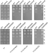
Sensitivity of M. alhagi CCNWXJ12-2T to different salts. Wild-type (WT), mutants (ΔrhcQ and ΔMA29250), and the complementation strains (CΔrhcQ and CΔMA29250) were grown to OD600 ≈0.8 in TY broth containing appropriate antibiotics, and then adjusted to OD600 ≈0.2. The inocula were serially diluted and spotted onto TY agar plates containing no additional salt or containing 0.4 M NaCl, 0.3 M LiCl, or 0.3 M KCl. The 10-fold serial dilutions are shown. All plates were incubated at 28°C for 4 days.
FIGURE 5
FIGURE 5
Total cellular Na+ and K+ concentrations of M. alhagi CCNWXJ12-2T. Wild-type (WT), mutants (ΔrhcQ and ΔMA29250), and the complementation strains (CΔrhcQ and CΔMA29250) were grown to OD600 ≈0.8 in TY broth containing appropriate antibiotics. A total of 500 microliters of each inoculum were plated on TY agar plates, TY plates containing 0.4 M NaCl, and TY plates containing 0.3 M KCl (all containing appropriate antibiotics) and inoculated at 28°C for 5 days. Then, the bacterial cells were collected for Na+ and K+ concentration measurement; those from TY plates containing 0.4 M NaCl were collected for Na+ concentration measurement, and those from TY plates containing 0.3 M KCl were collected for K+ concentration measurement. (A) Na+ concentrations in different strains; (B) K+ concentrations in different strains. Three independent experiments were conducted and the error bars indicate SDs. Significant differences between the WT strain and other strains are indicated (P < 0.05; ∗∗P < 0.01).
FIGURE 6
FIGURE 6
Survival rate of M. alhagi CCNWXJ12-2T under oxidative stress. Wild-type (WT), mutants (ΔrhcQ and ΔMA29250), and the complemented strains (CΔrhcQ and CΔMA29250) were grown to OD600 ≈0.8 in TY broth containing appropriate antibiotics, and then adjusted to OD600 ≈0.2. The cultures were treated with 10 mM H2O2 for 30 min or incubated without H2O2 as a control. Cell survival rates were then counted and compared to untreated cells. The mean values of three independent experiments are shown and the error bars indicate SDs. Significant differences between the WT strain and other strains are indicated (∗∗P < 0.01).
FIGURE 7
FIGURE 7
Antioxidant enzyme activities of M. alhagi CCNWXJ12-2T under oxidative and salt stress. Catalase (CAT) and superoxide dismutase (SOD) activities of the wild-type (WT), mutants (ΔrhcQ and ΔMA29250), and complemented strains (CΔrhcQ and CΔMA29250) were measured in three independent biological experiments. (A) CAT activities; (B) SOD activities. Three biological replicates were conducted and the error bars indicate SDs. Significant differences of the enzyme activities in different conditions between the WT strain and other strains are indicated (P < 0.05; ∗∗P < 0.01).

Similar articles

Cited by

References

    1. Aspedon A., Palmer K., Whiteley M. (2006). Microarray analysis of the osmotic stress response in Pseudomonas aeruginosa. J. Bacteriol. 188 2721–2725. 10.1128/JB.188.7.2721-2725.2006 - DOI - PMC - PubMed
    1. Biemans-Oldehinkel E., Sal-Man N., Deng W., Foster L. J., Finlay B. B. (2011). Quantitative proteomic analysis reveals formation of an EscL-EscQ-EscN Type III complex in enteropathogenic Escherichia coli. J. Bacteriol. 193 5514–5519. 10.1128/JB.05235-11 - DOI - PMC - PubMed
    1. Buttner D., He S. Y. (2009). Type III protein secretion in plant pathogenic bacteria. Plant Physiol. 150 1656–1664. 10.1104/pp.109.139089 - DOI - PMC - PubMed
    1. Bzymek K. P., Hamaoka B. Y., Ghosh P. (2012). Two translation products of Yersinia yscQ assemble to form a complex essential to Type III secretion. Biochemistry 51 1669–1677. 10.1021/bi201792p - DOI - PMC - PubMed
    1. Calderon-Torres M., Castro D. E., Montero P., Pena A. (2011). DhARO4 induction and tyrosine nitration in response to reactive radicals generated by salt stress in Debaryomyces hansenii. Yeast 28 733–746. 10.1002/yea.1903 - DOI - PubMed