

Improved Immune Responses in Young and Aged Mice with Adjuvanted Vaccines against H1N1 Influenza Infection
- PMID: 29515589
- PMCID: PMC5826078
- DOI: 10.3389/fimmu.2018.00295
Improved Immune Responses in Young and Aged Mice with Adjuvanted Vaccines against H1N1 Influenza Infection
Abstract
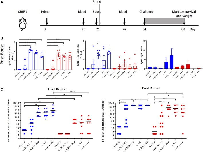
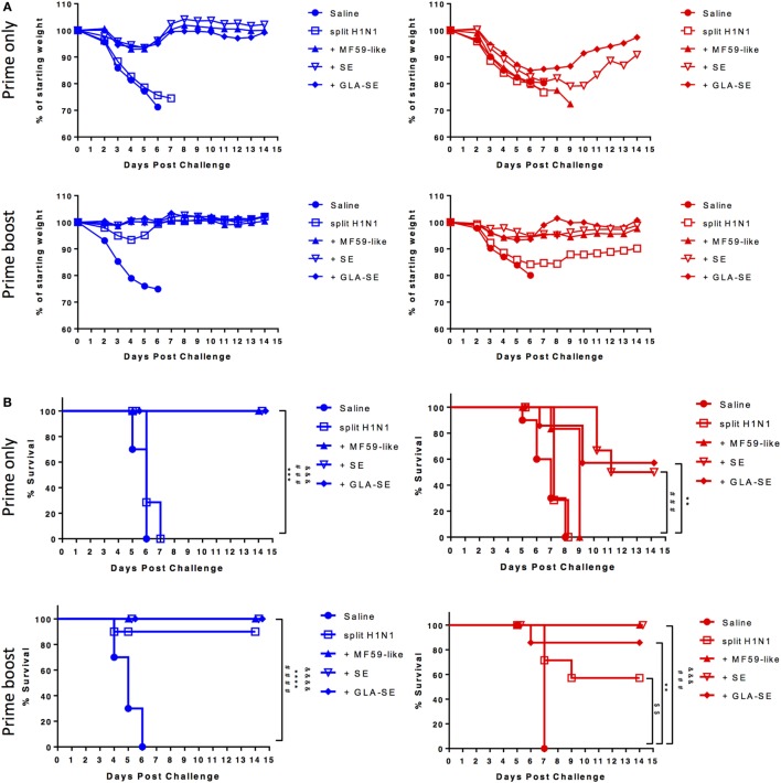
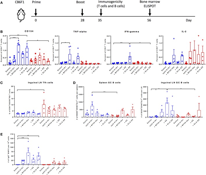
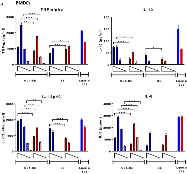
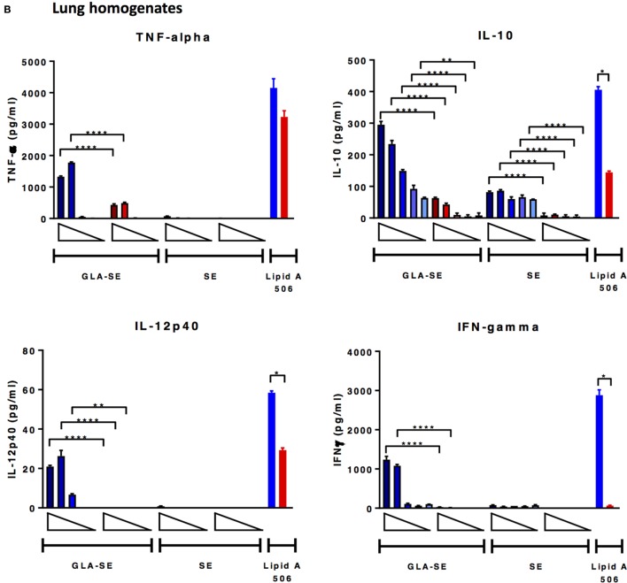
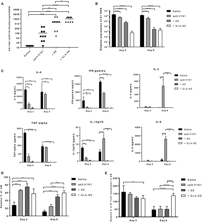
Elderly people are at high risk for influenza-related morbidity and mortality due to progressive immunosenescence. While toll-like receptor (TLR) agonist containing adjuvants, and other adjuvants, have been shown to enhance influenza vaccine-induced protective responses, the mechanisms underlying how these adjuvanted vaccines could benefit the elderly remain elusive. Here, we show that a split H1N1 influenza vaccine (sH1N1) combined with a TLR4 agonist, glucopyranosyl lipid adjuvant formulated in a stable oil-in-water emulsion (GLA-SE), boosts IgG2c:IgG1 ratios, enhances hemagglutination inhibition (HAI) titers, and increases protection in aged mice. We find that all adjuvanted sH1N1 vaccines tested were able to protect both young and aged mice from lethal A/H1N1/California/4/2009 virus challenge after two immunizations compared to vaccine alone. We show that GLA-SE combined with sH1N1, however, also provides enhanced protection from morbidity in aged mice given one immunization (based on change in weight percentage). While the GLA-SE-adjuvanted sH1N1 vaccine promotes the generation of cytokine-producing T helper 1 cells, germinal center B cells, and long-lived bone marrow plasma cells in young mice, these responses were muted in aged mice. Differential in vitro responses, dependent on age, were also observed from mouse-derived bone marrow-derived dendritic cells and lung homogenates following stimulation with adjuvants, including GLA-SE. Besides enhanced HAI titers, additional protective factors elicited with sH1N1 + GLA-SE in young mice were observed, including (a) rapid reduction of viral titers in the lung, (b) prevention of excessive lung inflammation, and (c) homeostatic maintenance of alveolar macrophages (AMs) following H1N1 infection. Collectively, our results provide insight into mechanisms of adjuvant-mediated immune protection in the young and elderly.
Keywords: H1N1; T helper 1; adjuvant; elderly; influenza; vaccine.
Figures
Similar articles
-
DAMP-Inducing Adjuvant and PAMP Adjuvants Parallelly Enhance Protective Type-2 and Type-1 Immune Responses to Influenza Split Vaccination.Front Immunol. 2018 Nov 20;9:2619. doi: 10.3389/fimmu.2018.02619. eCollection 2018. Front Immunol. 2018. PMID: 30515151 Free PMC article.
-
Overcoming the Neonatal Limitations of Inducing Germinal Centers through Liposome-Based Adjuvants Including C-Type Lectin Agonists Trehalose Dibehenate or Curdlan.Front Immunol. 2018 Feb 28;9:381. doi: 10.3389/fimmu.2018.00381. eCollection 2018. Front Immunol. 2018. PMID: 29541075 Free PMC article.
-
Synthetic Toll-like receptor 4 (TLR4) and TLR7 ligands as influenza virus vaccine adjuvants induce rapid, sustained, and broadly protective responses.J Virol. 2015 Mar;89(6):3221-35. doi: 10.1128/JVI.03337-14. Epub 2015 Jan 7. J Virol. 2015. PMID: 25568203 Free PMC article.
-
Inactivated influenza vaccines: recent progress and implications for the elderly.Drugs Aging. 2011 Feb 1;28(2):93-106. doi: 10.2165/11586770-000000000-00000. Drugs Aging. 2011. PMID: 21275435 Review.
-
Adjuvanted influenza vaccines.Hum Vaccin Immunother. 2018 Mar 4;14(3):550-564. doi: 10.1080/21645515.2017.1415684. Epub 2018 Jan 25. Hum Vaccin Immunother. 2018. PMID: 29232151 Free PMC article. Review.
Cited by
-
Host Factors Impact Vaccine Efficacy: Implications for Seasonal and Universal Influenza Vaccine Programs.J Virol. 2019 Oct 15;93(21):e00797-19. doi: 10.1128/JVI.00797-19. Print 2019 Nov 1. J Virol. 2019. PMID: 31391269 Free PMC article. Review.
-
Targeting TLR4 during vaccination boosts MAdCAM-1+ lymphoid stromal cell activation and promotes the aged germinal center response.Sci Immunol. 2022 May 6;7(71):eabk0018. doi: 10.1126/sciimmunol.abk0018. Epub 2022 May 6. Sci Immunol. 2022. PMID: 35522725 Free PMC article.
-
IgD+ age-associated B cells are the progenitors of the main T-independent B cell response to infection that generates protective Ab and can be induced by an inactivated vaccine in the aged.Aging Cell. 2022 Oct;21(10):e13705. doi: 10.1111/acel.13705. Epub 2022 Sep 2. Aging Cell. 2022. PMID: 36056604 Free PMC article.
-
Human Metapneumovirus Reinfection in Aged Mice Recapitulates Increased Disease Severity in Elderly Humans Infected with Human Metapneumovirus.Immunohorizons. 2023 Jun 1;7(6):398-411. doi: 10.4049/immunohorizons.2300026. Immunohorizons. 2023. PMID: 37261717 Free PMC article.
-
"World in motion" - emulsion adjuvants rising to meet the pandemic challenges.NPJ Vaccines. 2021 Dec 21;6(1):158. doi: 10.1038/s41541-021-00418-0. NPJ Vaccines. 2021. PMID: 34934069 Free PMC article. Review.
References
Publication types
MeSH terms
Substances
Grants and funding
LinkOut - more resources
Full Text Sources
Other Literature Sources
Medical