. 2018 Feb 7;5(2):171058.
doi: 10.1098/rsos.171058. eCollection 2018 Feb.

Identification of allosteric disulfides from labile bonds in X-ray structures

Affiliations

Identification of allosteric disulfides from labile bonds in X-ray structures

Aster E Pijning et al. R Soc Open Sci. .

Abstract

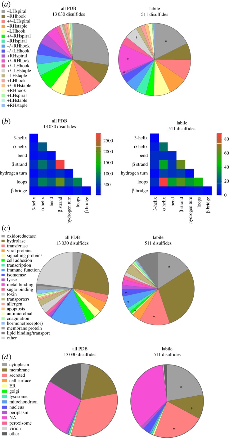
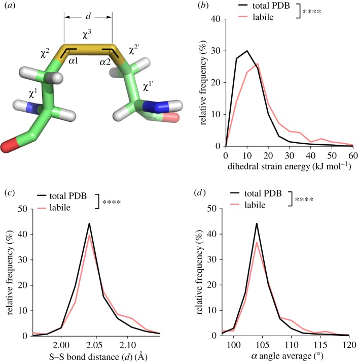
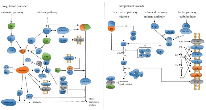
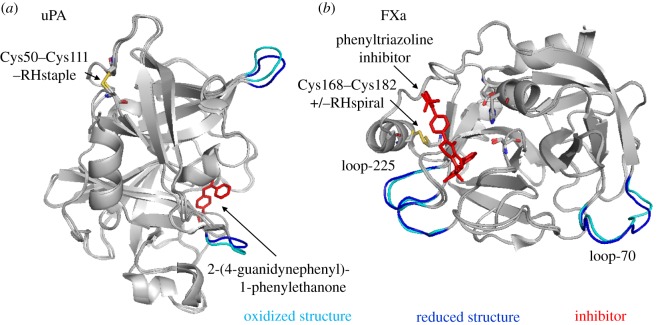
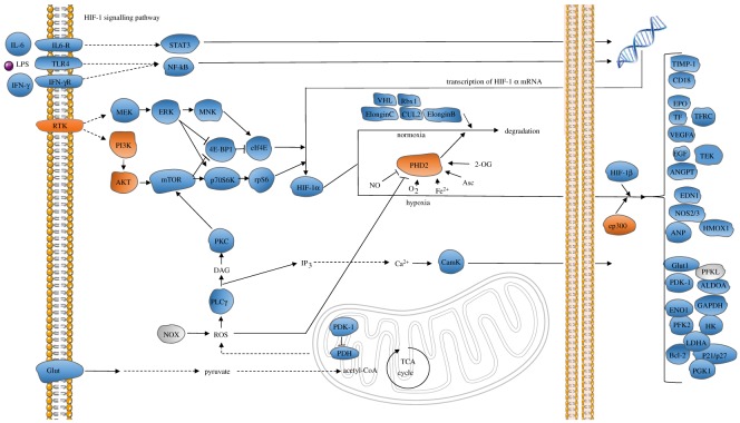
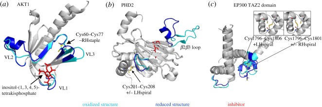
Protein disulfide bonds link pairs of cysteine sulfur atoms and are either structural or functional motifs. The allosteric disulfides control the function of the protein in which they reside when cleaved or formed. Here, we identify potential allosteric disulfides in all Protein Data Bank X-ray structures from bonds that are present in some molecules of a protein crystal but absent in others, or present in some structures of a protein but absent in others. We reasoned that the labile nature of these disulfides signifies a propensity for cleavage and so possible allosteric regulation of the protein in which the bond resides. A total of 511 labile disulfide bonds were identified. The labile disulfides are more stressed than the average bond, being characterized by high average torsional strain and stretching of the sulfur-sulfur bond and neighbouring bond angles. This pre-stress likely underpins their susceptibility to cleavage. The coagulation, complement and oxygen-sensing hypoxia inducible factor-1 pathways, which are known or have been suggested to be regulated by allosteric disulfides, are enriched in proteins containing labile disulfides. The identification of labile disulfide bonds will facilitate the study of this post-translational modification.

Keywords: allosteric disulfides; coagulation; complement; oxygen-sensing; redox.


Conflict of interest statement

We declare we have no competing interests.

Figures

Figure 1.
Figure 1.
Structural and functional features of the labile disulfide bonds. (a) Distribution of the 20 disulfide bond configurations in unique disulfide bonds in PDB protein X-ray structures (13 030 disulfides, electronic supplementary material, table S1) and in labile bonds (511 disulfides, electronic supplementary material, table S2). Compared with the total PDB, the labile bonds are enriched in +/–RHhook and +/–LHstaple bonds (χ2 test, p < 0.0001) and have relatively fewer –LHspiral and +RHspiral bonds (χ2 test, p < 0.0001) (indicated by *). (b) Heatmap displaying the frequency of the secondary structures linked by disulfide bonds in all PDB protein structures and by labile disulfide bonds. There is enrichment of disulfides linking α-helices and loops in labile disulfide bonds (χ2 test, p < 0.0001). (c) Distribution of the functional classification of all proteins containing disulfide bonds and proteins containing labile disulfide bonds. Compared to the total PDB, there was a significant increase in oxidoreductases, transferases and isomerases (indicated by *). A significant decrease in disulfide bonds in proteins involved in signalling and immune function was observed (χ2 test, p < 0.0001). (d) Subcellular localization of all proteins containing disulfide bonds and proteins containing labile disulfide bonds. Compared to the total PDB, a significant increase in cytoplasmic proteins, as well as a decrease in membrane associated and secreted proteins was observed (χ2 test, p < 0.0001) (indicated by *).
Figure 2.
Figure 2.
The labile disulfides are characterized by high dihedral strain energy, elongation of the sulfur–sulfur bond distance and stretching of the neighbouring bond angles. (a) Angles and distances of the cystine residue. The values α1 and α2 represent the two relevant bending angles of the disulfide, and the five dihedral angles are χ1, χ2, χ3, χ2′ and χ1′. d is the sulfur–sulfur bond length. (b) Relative frequency of DSE ranging from 0 to 60 kJ mol−1. The DSE was significantly increased for labile disulfides compared to all disulfide bonds in the PDB (p < 0.0001). (c) The relative frequency of the sulfur–sulfur bond distance ranging from 1.96 to 2.14 Å is shown. An increase in sulfur–sulfur bond distance is observed for labile disulfide bonds (p < 0.0001). (d) The average of both α angles was calculated for each disulfide bond. Shown is the relative frequency of the average angle ranging from 95 to 120°. Angles are increased for labile disulfide bonds (p < 0.0001). T-tests were used to compare total PDB to labile disulfides.
Figure 3.
Figure 3.
The coagulation and complement pathways are enriched in proteins containing labile disulfide bonds. The proteins containing labile disulfide bonds are shown in orange. The proteins shown in green contain characterized allosteric disulfide bonds (electronic supplementary material, table S3). Proteins shown in grey do not have any X-ray structures available in the PDB. FH: factor H. FI: complement factor I. FB: complement factor B. FD: complement factor D. C1NH: serpin family G member 1. C3: complement factor 3. C5: complement factor 5. DAF: CD55. MCP: CD46. CR1: complement C3b/C4b receptor 1. C1qrs: complement C1. MBL: mannose-binding lectin. MASP1/2: mannan-binding lectin serine peptidase 1/2. C2: complement C2. C4: complement C4. C4BP: complement component 4 binding protein. C5: complement C5. C6: complement C6. C7: complement C7. C8: complement C8. C9: complement C9. CLU: clusterin. VTN: vitronectin. C3AR1: complement C3a receptor 1. CRIg: V-set and immunoglobulin domain-containing protein 4. CR1: complement C3b/C4b receptor 1. CR2: complement C3d receptor 2. CR3: coagulation factor X/plasminogen. CR4: coagulation factor X/plasminogen. C5AR1: complement C5A receptor.
Figure 4.
Figure 4.
Conformational changes in proteins of the coagulation and complement pathways upon cleavage of the labile disulfide bonds. Structures of oxidized (cyan) and reduced (blue) proteins are aligned. Labile disulfides and their configurations are shown. The PDB identifiers are indicated in table 1. (a) uPA is shown in complex with the inhibitor, 2-(4-guanidynephenyl)-1-phenylethanone. The redox state of the Cys50–Cys111 disulfide influences the positions of loops surrounding the active site. (b) Factor Xa is shown in the presence of a phenyltriazoline inhibitor. The catalytic triad consisting of His57, Asp102 and Ser195 is shown in stick presentation. The redox state of Cys168–Cys182 influences slight conformational changes in Ca2+-binding loop-70 and Na2+-binding loop-225.
Figure 5.
Figure 5.
The HIF-1 signalling pathway is enriched in proteins containing labile disulfide bonds. The proteins containing labile disulfide bonds are shown in orange. Grey indicates that no X-ray structures are available of this protein. RTK: epidermal growth factor receptor/erb-b2 receptor tyrosine kinase. MEK: mitogen-activated protein kinase ½. ERK: mitogen-activated protein kinase 1/3. MNK: MAP kinase interacting kinase ½. STAT3: signal transducer and activator of transcription 3. 4E-BP1: eukaryotic translation initiation factor 4E-binding protein. eIf4E: eukaryotic translation initiation factor 4E. p70S6 K: ribosomal protein S6 kinase. Rps6: ribosomal protein S6. VHL: Von Hipper Lindau tumor suppressor. RBX1: ring box 1. CUL2: cullin 2. PI3 k: phosphoinositide-3-kinase. AKT: protein kinase B. mTOR: mechanistic target of rapamycin. ElonginC: transcription elongation factor B subunit 1. ElonginB: transcription elongation factor B subunit 2. PHD2: Egl-9 family hypoxia inducible factor 1. CamK: calcium/calmodium-dependent protein kinase. ep300: E1A-binding protein p300. PLCɣ: phospholipase C gamma. NOX: NADPH oxidase. Glut: solute carrier family 2 member 1. TIMP-1: TIMP metallopeptidase inhibitor 1. CD18: lymphotoxin beta receptor. EPO: erythropoietin. TF: transferrin. TFRC: transferrin receptor. VEGF: vascular endothelial growth factor. Flt-1: Fms-related tyrosine kinase 1. EGF: epidermal growth factor. TEK tyrosine Kinase: TIE-2, angiopoietin-1 receptor. EDN1: endothelin 1. NOS2/3: nitric oxide synthase 2/3. ANP: natriuretic peptide A. PDK-1: pyruvate dehydrogenase kinase 1. HK: hexokinase. PFKL: ATP-dependent 6-phosphofructokinase. GAPDH: glyceraldehyde-3-phosphate dehydrogenase. ALDOA: fructose-bisphosphate aldolase A. ENO1: enolase 1. PGK1: phosphoglycerate kinase 1. PFK2: 6-phosphofructo-2-kinase/fructose-2,6-biphosphatase 3. LDHA: l-lactate dehydrogenase A chain. Bcl-2: apoptosis regulator Bcl-2. p21/p27: cyclin-dependent kinase inhibitor 1.
Figure 6.
Figure 6.
Conformational changes in HIF-1 pathway proteins upon cleavage of the labile disulfide bonds. Structures of oxidized (cyan) and reduced (blue) proteins are aligned. Labile disulfides and their configurations are shown. The PDB identifiers are indicated in table 2. (a) PH domain of AKT1 with bound phosphoinositol. The positions of variable loops (VL) 1, 2 and 3 differ in oxidized and reduced structures. (b) The position of the β2β3 loop of PHD2 differs in oxidized and reduced structures. (c) The positions of the zinc-binding loops connecting the α-helices in the TAZ2 domain of EP300 differ in oxidized and reduced structures. The insets depict the two possibilities of disulfide bonding between the three participating cysteines within the oxidized structure.

Similar articles

Cited by

References

    1. Cook KM, Hogg PJ. 2013. Post-translational control of protein function by disulfide bond cleavage. Antioxid. Redox Signal. 18, 1987–2015. (doi:10.1089/ars.2012.4807) - DOI - PubMed
    1. Butera D, Cook KM, Chiu J, Wong JW, Hogg PJ. 2014. Control of blood proteins by functional disulfide bonds. Blood 123, 2000–2007. (doi:10.1182/blood-2014-01-549816) - DOI - PMC - PubMed
    1. Hogg PJ. 2013. Targeting allosteric disulphide bonds in cancer. Nat. Rev. Cancer 13, 425–431. (doi:10.1038/nrc3519) - DOI - PubMed
    1. Wouters MA, Lau KK, Hogg PJ. 2004. Cross-strand disulphides in cell entry proteins: poised to act. Bioessays 26, 73–79. (doi:10.1002/bies.10413) - DOI - PubMed
    1. Bekendam RH, et al. 2016. A substrate-driven allosteric switch that enhances PDI catalytic activity. Nat. Commun. 7, 12579 (doi:10.1038/ncomms12579) - DOI - PMC - PubMed