Clinical Trial
. 2018 Sep;45(10):1762-1771.
doi: 10.1007/s00259-018-3980-9. Epub 2018 Mar 8.

18F-Fluorocholine PET/CT in the assessment of primary hyperparathyroidism compared with 99mTc-MIBI or 99mTc-tetrofosmin SPECT/CT: a prospective dual-centre study in 100 patients

Affiliations
Clinical Trial

18F-Fluorocholine PET/CT in the assessment of primary hyperparathyroidism compared with 99mTc-MIBI or 99mTc-tetrofosmin SPECT/CT: a prospective dual-centre study in 100 patients

Mohsen Beheshti et al. Eur J Nucl Med Mol Imaging. 2018 Sep.

Abstract

Purpose: In this prospective study we compared the accuracy of 18F-fluorocholine PET/CT with that of 99mTc-MIBI or99mTc-tetrofosmin SPECT/CT in the preoperative detection of parathyroid adenoma in patients with primary hyperparathyroidism. We also assessed the value of semiquantitative parameters in differentiating between parathyroid hyperplasia and adenoma.

Methods: Both 18F-fluorocholine PET/CT and 99mTc-MIBI/tetrofosmin SPECT/CT were performed in 100 consecutive patients with biochemical evidence of primary hyperparathyroidism. At least one abnormal focus on either 18F-fluorocholine or 99mTc-MIBI/tetrofosmin corresponding to a parathyroid gland or ectopic parathyroid tissue was considered as a positive finding. In 76 patients with positive findings on at least one imaging modality, surgical exploration was performed within 6 months, and the results were related to histopathological findings and clinical and laboratory findings at 3-6 months as the standard of truth. In 24 patients, no surgery was performed: in 18 patients with positive imaging findings surgery was refused or considered risky, and in 6 patients imaging was negative. Therefore, data from 82 patients (76 undergoing surgery, 6 without surgery) in whom the standard of truth criteria were met, were used in the final analysis.

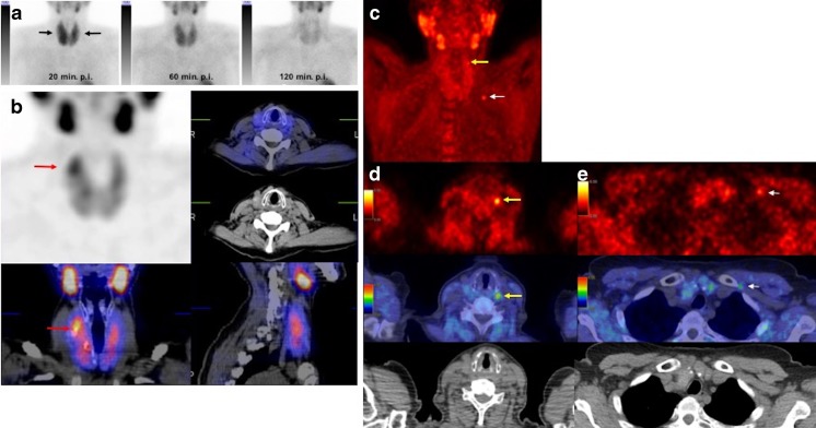
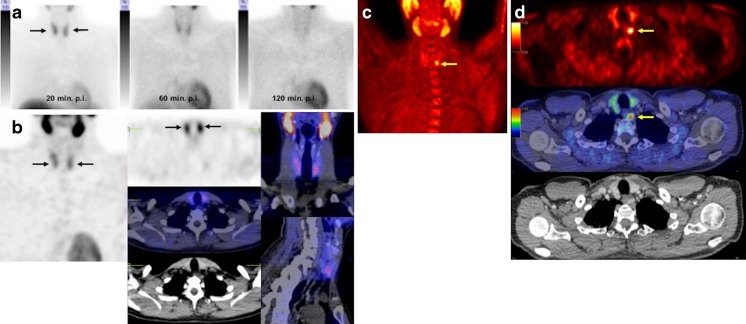
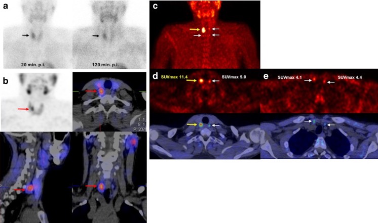
Results: All patients showed biochemical evidence of primary hyperparathyroidism with a mean serum calcium level of 2.78 ± 0.34 mmol/l and parathormone (PTH) level of 196.5 ± 236.4 pg/ml. The study results in 76 patients with verified histopathology and 3 patients with negative imaging findings were analysed. Three of six patients with negative imaging showed normalized serum PTH and calcium levels on laboratory follow-up at 3 and 6 months, and the results were considered true negative. In a patient-based analysis, the detection rate with 18F-fluorocholine PET/CT was 93% (76/82), but was only 61% (50/82) with 99mTc-MIBI/tetrofosmin SPECT/CT. In a lesion-based analysis, the sensitivity, specificity, positive predictive value, negative predictive value and overall accuracy of 18F-fluorocholine PET/CT in the detection of parathyroid adenoma were 93.7%, 96.0%, 90.2%, 97.4% and 95.3%, respectively, and of 99mTc-MIBI/tetrofosmin SPECT/CT were 60.8%, 98.5%, 94.1%, 86.3% and 87.7%, respectively. Although 18F-fluorocholine PET-positive adenomatous lesions showed higher SUVmax values than the hyperplastic glands (6.80 ± 3.78 vs. 4.53 ± 0.40) in the semiquantitative analysis, the difference was not significant (p = 0.236). The mean size (measured as the length of the greatest dimension) and weight of adenomas were 15.9 ± 7.6 mm (median 15 mm, range 1-40 mm) and 1.71 ± 1.86 g (median 1 g, range: 0.25-9 g), respectively. Among the analysed parameters including serum calcium and PTH and the size and weight of parathyroid adenomas, size was significantly different between patients with negative 99mTc-MIBI/tetrofosmin SPECT/CT and those with positive 99mTc-MIBI/tetrofosmin SPECT/CT (mean size 13.4 ± 7.6 mm vs. 16.9 ± 7.4 mm, respectively; p = 0.042).

Conclusion: In this prospective study, 18F-fluorocholine PET/CT showed promise as a functional imaging modality, being clearly superior to 99mTc-MIBI/tetrofosmin SPECT/CT, especially in the detection and localization of small parathyroid adenomas in patients with primary hyperparathyroidism. SUVmax was higher in parathyroid adenomas than in hyperplasia. However, further evaluation of this modality is needed.

Keywords: 18F-Fluorocholine PET/CT; 99mTc-MIBI; 99mTc-Tetrofosmin; Primary hyperparathyroidism; SPECT/CT.


Conflict of interest statement

Conflicts of interest

None.

Ethical approval

All procedures performed in studies involving human participants were in accordance with the ethical standards of the institutional and/or national research committee and with the principles of the 1964 Declaration of Helsinki and its later amendments or comparable ethical standards.

Figures

Fig. 1
Fig. 1
99mTc-MIBI planar scintigraphy images (a), 99mTc-MIBI SPECT/CT images (b) and 18F-fluorocholine PET/CT images (c–e) in a 48-year-old patient with elevated serum PTH and calcium levels of 91.29 pg/ml and 2.87 mmol/l, respectively. a Anterior 99mTc-MIBI planar images show diffuse nonhomogeneous tracer uptake in the enlarged thyroid gland in the image 20 min after injection (20 min p.i. arrows) with regular wash-in seen on the 60-min image (60 min p.i.) and 120-min image (120 min p.i.) without any evidence of focal tracer retention suggestive of parathyroid adenoma. b 99mTc-MIBI SPECT/CT images (60 min after injection) also show no evidence of pathological uptake suggestive of parathyroid adenoma. The focal tracer uptake in the upper part of the right thyroid is suggestive of thyroid adenoma (arrows). c–e 18F-Fluorocholine PET/CT images: maximum intensity projection (MIP) image (c) and transaxial PET images (d top), PET/CT fusion images (d middle) and CT images (d bottom) show marked focal tracer uptake in the upper part of the left thyroid suggestive of parathyroid adenoma (yellow arrows) which was confirmed on histopathology (size 9 mm, weight <1 g). Focal tracer uptake is seen in the left clavicular region on the MIP image (c white arrow) which is confirmed as nonspecific vascular uptake on the delayed images (e arrows)
Fig. 2
Fig. 2
99mTc-MIBI planar scintigraphy images (a), 99mTc-MIBI SPECT/CT images (b) and 18F-fluorocholine PET/CT images (c, d) in a 67-year-old woman with an elevated serum PTH level of 108.4 pg/ml and a borderline serum calcium level of 2.51 mmol/l. a Anterior 99mTc-MIBI planar images show homogeneous tracer uptake in the thyroid gland in the image 20 min after injection (20 min p.i. arrows) with regular wash-in seen on the 60-min image (60 min p.i.) and 120-min image (120 min p.i.) without any evidence of focal tracer retention suggestive of parathyroid adenoma. b 99mTc-MIBI SPECT/CT images (60 min after injection) also show homogeneous tracer uptake in the thyroid gland (arrows), without any evidence of pathological uptake suggestive of parathyroid adenoma. c, d 18F-Fluorocholine PET/CT images: maximum intensity projection (MIP) image (c) and transaxial PET image (d top), PET/CT fusion image (d middle) and CT image (d bottom) show marked focal tracer uptake in the lower part of the left thyroid suggestive of parathyroid adenoma (arrows)
Fig. 3
Fig. 3
99mTc-MIBI planar scintigraphy images (a), 99mTc-MIBI SPECT/CT images (b) and 18F-fluorocholine PET/CT images (c–e) in a 35-year-old man with elevated serum PTH and calcium levels of 512.0 pg/ml and 2.82 mmol/l, respectively. a Anterior 99mTc-MIBI planar images show nonhomogeneous tracer uptake in the thyroid glands with focal uptake on the right side in the image 20 min after injection (20 min p.i. arrows) with irregular wash-in in the 120-min image (120 min p.i.) and evidence of focal tracer retention suggestive of thyroid or parathyroid adenoma. b 99mTc-tetrofosmin SPECT/CT images (120 min after injection) show pathological focal uptake in the posterior part of the right thyroid lobe suggestive of parathyroid adenoma. c–e 18F-Fluorocholine PET/CT images: maximum intensity projection (MIP) image (c) shows focal tracer uptake in the upper posterior part of the right thyroid lobe (yellow arrows) with three small areas of mild tracer uptake in the other parathyroid glands (white arrows) which was found to be hyperplastic parathyroid glands on histopathology; d transaxial PET image (top) and PET/CT fusion image (bottom) show marked focal tracer uptake in the upper part of the right thyroid suggestive of parathyroid adenoma (yellow arrows) and a small mild focal lesion in the upper part of the left thyroid suggestive of parathyroid hyperplasia (white arrows); e transaxial PET image (top) and PET/CT fusion image (bottom) show marked small mild focal lesions on the lower part of both thyroid lobes suggestive of parathyroid hyperplasia

References

    1. Taniegra ED. Hyperparathyroidism. Am Fam Physician. 2004;69:333–339. - PubMed
    1. Madkhali T, Alhefdhi A, Chen H, Elfenbein D. Primary hyperparathyroidism. Ulus Cerrahi Derg. 2016;32:58–66. - PMC - PubMed
    1. Taieb D, Hindie E, Grassetto G, Colletti PM, Rubello D. Parathyroid scintigraphy: when, how, and why? A concise systematic review. Clin Nucl Med. 2012;37:568–574. doi: 10.1097/RLU.0b013e318251e408. - DOI - PubMed
    1. Wilhelm SM, Wang TS, Ruan DT, Lee JA, Asa SL, Duh QY, et al. The American association of endocrine surgeons guidelines for definitive management of primary hyperparathyroidism. JAMA Surg. 2016;151:959–968. doi: 10.1001/jamasurg.2016.2310. - DOI - PubMed
    1. Hocevar M, Lezaic L, Rep S, Zaletel K, Kocjan T, Sever MJ, et al. Focused parathyroidectomy without intraoperative parathormone testing is safe after pre-operative localization with 18F-fluorocholine PET/CT. Eur J Surg Oncol. 2017;43:133–137. doi: 10.1016/j.ejso.2016.09.016. - DOI - PubMed