

Malignant atrophic papulosis with motor aphasia and intestinal perforation: A case report and review of published works
- PMID: 29516548
- PMCID: PMC6001538
- DOI: 10.1111/1346-8138.14280
Malignant atrophic papulosis with motor aphasia and intestinal perforation: A case report and review of published works
Abstract
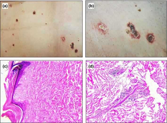
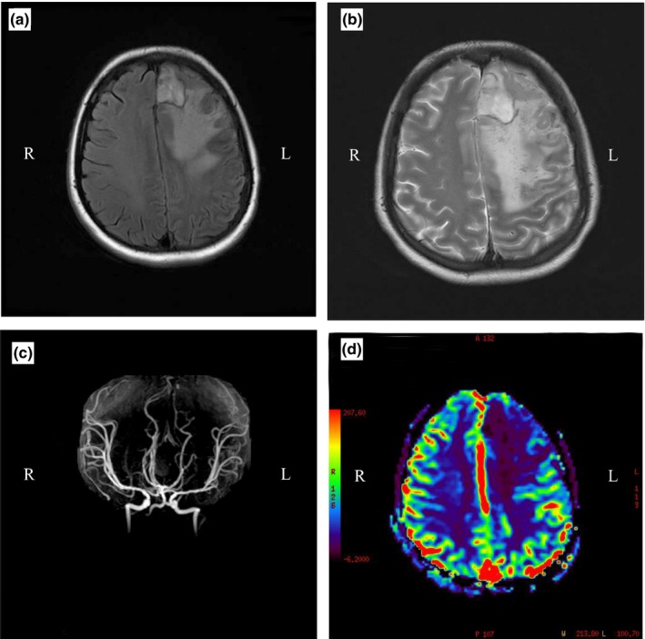
Malignant atrophic papulosis (MAP) is a rare type of obliterating vasculopathy that can present as pure cutaneous lesions or a systemic entity affecting multiple organs. Systemic disease, such as gastrointestinal or central nervous system involvement, may predispose the patients to poorer or even fatal outcomes. We present a 30-year-old female patient with systemic manifestation of MAP 10 days after delivery of a full-term pregnancy who subsequently developed motor aphasia and intestinal perforation. The patient was administrated empirical treatment with an antiplatelet, anticoagulant, methylprednisolone sodium succinate and alprostadil. Antibiotics were administrated due to intestinal perforation and secondary sepsis. Despite all treatment, the patient died a week later. We summarized all the previous reports of MAP based on thorough review of previous published work. Overall, this is the first patient with MAP combined with motor aphasia and intestinal perforation and may provide insights for future studies on the treatment of this disease.
Keywords: case report; intestinal perforation; malignant atrophic papulosis; motor aphasia; review of published work.
© 2018 The Authors. The Journal of Dermatology published by John Wiley & Sons Australia, Ltd on behalf of Japanese Dermatological Association.
Figures
References
-
- Köhlmeier W. Multiple Hautnekrosen bei Thrombangiitis obliterans. Arch Dermatol Syph 1941; 181: 783–792.
-
- Scheinfeld N. Malignant atrophic papulosis. Clin Exp Dermatol 2007; 32: 483–487. - PubMed
-
- Heymann WR. Degos disease: considerations for reclassification. J Am Acad Dermatol 2009; 61: 505–506. - PubMed
-
- Theodoridis A, Konstantinidou A, Makrantonaki E, Zouboulis CC. Malignant and benign forms of atrophic papulosis (Kohlmeier‐Degos disease): systemic involvement determines the prognosis. Br J Dermatol 2014; 170: 110–115. - PubMed
Publication types
MeSH terms
Substances
LinkOut - more resources
Full Text Sources
Other Literature Sources
Medical
