Review
. 2018 Jan 1;9(1):216-226.
doi: 10.1080/19491034.2018.1449498.

Laminopathy-causing lamin A mutations reconfigure lamina-associated domains and local spatial chromatin conformation

Affiliations
Review

Laminopathy-causing lamin A mutations reconfigure lamina-associated domains and local spatial chromatin conformation

Nolwenn Briand et al. Nucleus. .

Abstract

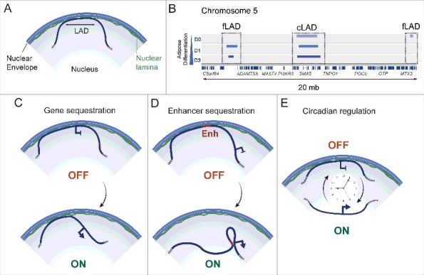
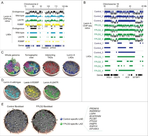
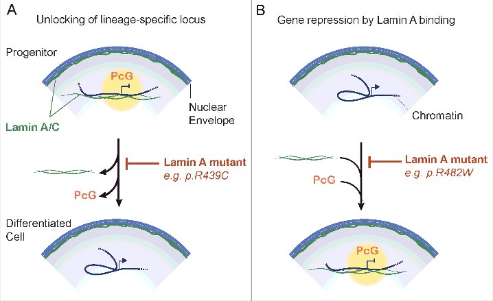
The nuclear lamina contributes to the regulation of gene expression and to chromatin organization. Mutations in A-type nuclear lamins cause laminopathies, some of which are associated with a loss of heterochromatin at the nuclear periphery. Until recently however, little if any information has been provided on where and how lamin A interacts with the genome and on how disease-causing lamin A mutations may rearrange genome conformation. Here, we review aspects of nuclear lamin association with the genome. We highlight recent evidence of reorganization of lamin A-chromatin interactions in cellular models of laminopathies, and implications on the 3-dimensional rearrangement of chromatin in these models, including patient cells. We discuss how a hot-spot lipodystrophic lamin A mutation alters chromatin conformation and epigenetic patterns at an anti-adipogenic locus, and conclude with remarks on links between lamin A, Polycomb and the pathophysiology of laminopathies. The recent findings presented here collectively argue towards a deregulation of large-scale and local spatial genome organization by a subset of lamin A mutations causing laminopathies.

Keywords: 3D genome; LAD; chromatin; differentiation; genome conformation; lamin A/C.


Figures

Figure 1.
Figure 1.
Modes of regulation of gene expression by nuclear lamins. (A) A lamina-associated domain (LAD). (B) Genome browser view of lamin A LADs during adipose differentiation of human adipocyte progenitors into adipocytes (day 0, 1, 3 of differentiation). Facultative fLADs and constitutive cLADs are shown in a region of chromosome 5. LAD data are from reference [31]. (C-E) Regulation of developmental gene expression by (C) sequestration/release of a whole gene locus at/from the nuclear envelope, (D) sequestration/release of an enhancer at/from the nuclear envelope and (E) circadian association a gene locus with, and release from, the nuclear envelope.
Figure 2.
Figure 2.
3D genome modeling provides a spatial appreciation of LADs altered by lamin A mutations causing laminopathies. (A) Genome browser view of lamin A ChIP-seq profiles and LADs in a region of chromosome 2 in HeLa cells expressing indicated lamins. (B) Lamin A ChIP-seq profiles and LADs in a region of chromosome 18, in three control and four FPLD2 patient fibroblast cultures, the latter all with the R482W mutation. LAD data in (A) and (B) are from reference [26]. (C) A 3D structural model of HeLa cell nuclei. The whole-genome model reflects chromosome territories; each chromosome is modeled as a chain of beads representing TADs, and is colored differently. Chromosome territories are also visible in the tomographic view. Tomographic views of the same genome structure also reveal TADs interacting pairwise (red) and TADs containing LADs (blue) (adapted from reference [26). (D) 3D genome models showing LADs generated by expression of indicated lamins in HeLa cells [26]. (E) 3D genome models showing LADs specific to control and FPLD2 fibroblasts with the lamin A R482W mutation (from reference [26). Box, genes pertaining to white and brown adipogenesis found in FLPD2-specific LADs. Nucleus radius in the models in (C-E) is 5 µm. All panels were from reference [26] and were used with permission.
Figure 3.
Figure 3.
Connecting lamin A mutations, Polycomb and local genome conformation to developmental gene expression. (A) Unlocking of a lineage-specific gene from lamin A. In progenitor cells, a locus is held in a repressed state by Polycomb proteins (PcG) stabilized intranuclear lamin A/C. Dissociation of lamin A/C from the locus upon differentiation favors release of Polycomb, promoter-enhancer interaction and transcriptional activation of the locus. Some lamin A mutants such as lamin A p.R439C [72] enhance lamin A binding to chromatin and would inhibit this process. (B) Conversely, a gene active in progenitor cells is repressed by Polycomb on differentiation. This is enabled by an intranuclear lamin A network which stabilizes Polycomb at the locus. An example is the MIR335 gene in adipocyte progenitors [22]. The lamin A p.R482W mutant prevents lamin A binding, Polycomb recruitment and transcriptional repression.

References

    1. Burke B, Stewart CL. The nuclear lamins: flexibility in function. Nat Rev Mol Cell Biol. 2013;14:13–24. - PubMed
    1. Collas P, Lund EG, Oldenburg AR. Closing the (nuclear) envelope on the genome: how nuclear lamins interact with promoters and modulate gene expression. BioEssays. 2014;36:75–83. - PubMed
    1. van Steensel B Belmont AS. Lamina-Associated Domains: Links with Chromosome Architecture, Heterochromatin, and Gene Repression. Cell. 2017;169:780–791. - PMC - PubMed
    1. Kolb T, Maass K, Hergt M, et al.. Lamin A and lamin C form homodimers and coexist in higher complex forms both in the nucleoplasmic fraction and in the lamina of cultured human cells. Nucleus. 2011;2:425–433. - PubMed
    1. Lund E, Oldenburg A, Delbarre E, et al.. Lamin A/C-promoter interactions specify chromatin state-dependent transcription outcomes. Genome Res. 2013;23:1580–1589. - PMC - PubMed

Publication types