

Fish polyomaviruses belong to two distinct evolutionary lineages
- PMID: 29517483
- PMCID: PMC5982132
- DOI: 10.1099/jgv.0.001041
Fish polyomaviruses belong to two distinct evolutionary lineages
Abstract
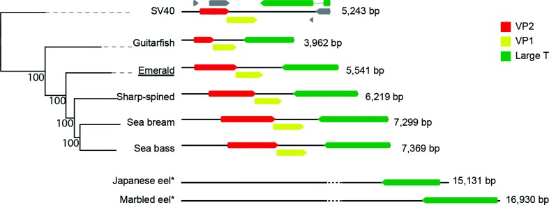
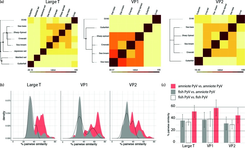
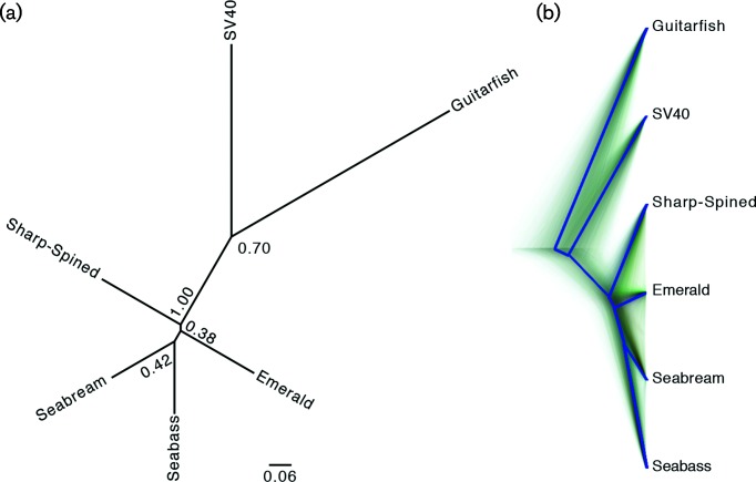
The Polyomaviridae is a diverse family of circular double-stranded DNA viruses. Polyomaviruses have been isolated from a wide array of animal hosts. An understanding of the evolutionary and ecological dynamics of these viruses is essential to understanding the pathogenicity of polyomaviruses. Using a high throughput sequencing approach, we identified a novel polyomavirus in an emerald notothen (Trematomus bernacchii) sampled in the Ross sea (Antarctica), expanding the known number of fish-associated polyomaviruses. Our analysis suggests that polyomaviruses belong to three main evolutionary clades; the first clade is made up of all recognized terrestrial polyomaviruses. The fish-associated polyomaviruses are not monophyletic, and belong to two divergent evolutionary lineages. The fish viruses provide evidence that the evolution of the key viral large T protein involves gain and loss of distinct domains.
Keywords: Antarctica; Polyomaviridae; Trematomus bernacchii; emerald notothen.
Conflict of interest statement
The authors declare that there are no conflicts of interest.
Figures

Similar articles
-
Identification of a polyomavirus in Weddell seal (Leptonychotes weddellii) from the Ross Sea (Antarctica).Arch Virol. 2017 May;162(5):1403-1407. doi: 10.1007/s00705-017-3239-y. Epub 2017 Jan 25. Arch Virol. 2017. PMID: 28124141
-
Discovery of African bat polyomaviruses and infrequent recombination in the large T antigen in the Polyomaviridae.J Gen Virol. 2017 Apr;98(4):726-738. doi: 10.1099/jgv.0.000737. Epub 2017 Apr 22. J Gen Virol. 2017. PMID: 28430100
-
Characterization of two novel polyomaviruses of birds by using multiply primed rolling-circle amplification of their genomes.J Virol. 2006 Apr;80(7):3523-31. doi: 10.1128/JVI.80.7.3523-3531.2006. J Virol. 2006. PMID: 16537620 Free PMC article.
-
Genome analysis of non-human primate polyomaviruses.Infect Genet Evol. 2014 Aug;26:283-94. doi: 10.1016/j.meegid.2014.05.030. Epub 2014 Jun 14. Infect Genet Evol. 2014. PMID: 24933462 Review.
-
[New, newer, newest human polyomaviruses: how far?].Mikrobiyol Bul. 2013 Apr;47(2):362-81. doi: 10.5578/mb.5377. Mikrobiyol Bul. 2013. PMID: 23621738 Review. Turkish.
Cited by
-
A novel lineage of polyomaviruses identified in bark scorpions.Virology. 2021 Nov;563:58-63. doi: 10.1016/j.virol.2021.08.008. Epub 2021 Aug 18. Virology. 2021. PMID: 34425496 Free PMC article.
-
A parasite outbreak in notothenioid fish in an Antarctic fjord.iScience. 2022 Jun 15;25(7):104588. doi: 10.1016/j.isci.2022.104588. eCollection 2022 Jul 15. iScience. 2022. PMID: 35800770 Free PMC article.
-
Identification of a novel polyomavirus in wild Sonoran Desert rodents of the family Heteromyidae.Arch Virol. 2023 Sep 15;168(10):253. doi: 10.1007/s00705-023-05877-5. Arch Virol. 2023. PMID: 37715108
-
Novel Polyomaviruses in Mammals from Multiple Orders and Reassessment of Polyomavirus Evolution and Taxonomy.Viruses. 2019 Oct 10;11(10):930. doi: 10.3390/v11100930. Viruses. 2019. PMID: 31658738 Free PMC article.
-
Diverse Microvirus Genomes Identified in the Stomach of a Sharp-Spined Notothen (Trematomus pennellii) from the Ross Sea (East Antarctica).Microbiol Resour Announc. 2023 Feb 16;12(2):e0123322. doi: 10.1128/mra.01233-22. Epub 2023 Jan 26. Microbiol Resour Announc. 2023. PMID: 36700633 Free PMC article.
References
-
- Gross L. The fortuitous isolation and identification of the polyoma virus. Cancer Res. 1976;36:4195–4196. - PubMed
Publication types
MeSH terms
Substances
LinkOut - more resources
Full Text Sources
Other Literature Sources