Magnetic susceptibility increases as diamagnetic molecules breakdown: Myelin digestion during multiple sclerosis lesion formation contributes to increase on QSM
- PMID: 29517817
- PMCID: PMC6129234
- DOI: 10.1002/jmri.25997
Magnetic susceptibility increases as diamagnetic molecules breakdown: Myelin digestion during multiple sclerosis lesion formation contributes to increase on QSM
Abstract
Background: The pathological processes in the first weeks of multiple sclerosis (MS) lesion formation include myelin digestion that breaks chemical bonds in myelin lipid layers. This can increase lesion magnetic susceptibility, which is a potentially useful biomarker in MS patient management, but not yet investigated.
Purpose: To understand and quantify the effects of myelin digestion on quantitative susceptibility mapping (QSM) of MS lesions.
Study type: Histological and QSM analyses on in vitro models of myelin breakdown and MS lesion formation in vivo.
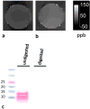
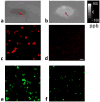
Population/specimens: Acutely demyelinating white matter lesions from MS autopsy tissue were stained with the lipid dye oil red O. Myelin basic protein (MBP), a major membrane protein of myelin, was digested with trypsin. Purified human myelin was denatured with sodium dodecyl sulfate (SDS). QSM was performed on phantoms containing digestion products and untreated controls. In vivo QSM was performed on five MS patients with newly enhancing lesions, and then repeated within 2 weeks.
Field strength/sequence: 3D -weighted spoiled multiecho gradient echo scans performed at 3T.
Assessment: Region of interest analyses were performed by a biochemist and a neuroradiologist to determine susceptibility changes on in vitro and in vivo QSM images.
Statistical tests: Not applicable.
Results: MBP degradation by trypsin increased the QSM measurement by an average of 112 ± 37 ppb, in excellent agreement with a theoretical estimate of 111 ppb. Degradation of human myelin by SDS increased the QSM measurement by 23 ppb. As MS lesions changed from gadolinium enhancing to nonenhancing over an average of 15.8 ± 3.7 days, their susceptibility increased by an average of 7.5 ± 6.3 ppb.
Data conclusion: Myelin digestion in the early stages of MS lesion formation contributes to an increase in tissue susceptibility, detectable by QSM, as a lesion evolves from gadolinium enhancing to nonenhancing.
Level of evidence: 1 Technical Efficacy: Stage 3 J. Magn. Reson. Imaging 2018;47:1281-1287.
Keywords: QSM; diamagnetism; multiple sclerosis; myelin.
© 2018 International Society for Magnetic Resonance in Medicine.
Figures


References
-
- Kutzelnigg A, Lassmann H. Pathology of multiple sclerosis and related inflammatory demyelinating diseases. Handb Clin Neurol. 2014;122:15–58. - PubMed
-
- Greenfield JG, Love S, Louis DN, Ellison D. Greenfield's neuropathology. London: Hodder Arnold; 2008.
-
- Kuhlmann T, Ludwin S, Prat A, Antel J, Brück W, Lassmann H. An updated histological classification system for multiple sclerosis lesions. Acta Neuropathol. 2017;133(1):13–24. - PubMed
Publication types
MeSH terms
Substances
Grants and funding
LinkOut - more resources
Full Text Sources
Other Literature Sources
Medical
Research Materials
Miscellaneous

