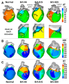
Multi-Scale Assessments of Cardiac Electrophysiology Reveal Regional Heterogeneity in Health and Disease
- PMID: 29517992
- PMCID: PMC5872364
- DOI: 10.3390/jcdd5010016
Multi-Scale Assessments of Cardiac Electrophysiology Reveal Regional Heterogeneity in Health and Disease
Abstract
The left and right ventricles of the four-chambered heart have distinct developmental origins and functions. Chamber-specific developmental programming underlies the differential gene expression of ion channel subunits regulating cardiac electrophysiology that persists into adulthood. Here, we discuss regional specific electrical responses to genetic mutations and cardiac stressors, their clinical correlations, and describe many of the multi-scale techniques commonly used to analyze electrophysiological regional heterogeneity.
Keywords: ECGI; cardiac development; electrophysiology; optical mapping.
Conflict of interest statement
The authors declare no conflict of interest.
Figures


Similar articles
-
Differential Wnt-mediated programming and arrhythmogenesis in right versus left ventricles.J Mol Cell Cardiol. 2018 Oct;123:92-107. doi: 10.1016/j.yjmcc.2018.09.002. Epub 2018 Sep 5. J Mol Cell Cardiol. 2018. PMID: 30193957 Free PMC article.
-
Quantitative reconstruction of cardiac electromechanics in human myocardium: regional heterogeneity.J Cardiovasc Electrophysiol. 2003 Oct;14(10 Suppl):S219-28. doi: 10.1046/j.1540.8167.90314.x. J Cardiovasc Electrophysiol. 2003. PMID: 14760927
-
Differences in Left Versus Right Ventricular Electrophysiological Properties in Cardiac Dysfunction and Arrhythmogenesis.Arrhythm Electrophysiol Rev. 2016 May;5(1):14-9. doi: 10.15420/aer.2016.8.2. Arrhythm Electrophysiol Rev. 2016. PMID: 27403288 Free PMC article.
-
Optical and electrical recordings from isolated coronary-perfused ventricular wedge preparations.J Mol Cell Cardiol. 2013 Jan;54:53-64. doi: 10.1016/j.yjmcc.2012.10.017. Epub 2012 Nov 9. J Mol Cell Cardiol. 2013. PMID: 23142540 Free PMC article. Review.
-
Differential distribution of cardiac ion channel expression as a basis for regional specialization in electrical function.Circ Res. 2002 May 17;90(9):939-50. doi: 10.1161/01.res.0000018627.89528.6f. Circ Res. 2002. PMID: 12016259 Review.
Cited by
-
Proceedings From the 2019 Stanford Single Ventricle Scientific Summit: Advancing Science for Single Ventricle Patients: From Discovery to Clinical Applications.J Am Heart Assoc. 2020 Apr 7;9(7):e015871. doi: 10.1161/JAHA.119.015871. Epub 2020 Mar 19. J Am Heart Assoc. 2020. PMID: 32188306 Free PMC article.
References
-
- Sivagangabalan G., Nazzari H., Bignolais O., Maguy A., Naud P., Farid T., Masse S., Gaborit N., Varro A., Nair K., et al. Regional ion channel gene expression heterogeneity and ventricular fibrillation dynamics in human hearts. PLoS ONE. 2014;9:e82179. doi: 10.1371/journal.pone.0082179. - DOI - PMC - PubMed
Publication types
Grants and funding
LinkOut - more resources
Full Text Sources
Other Literature Sources

