

Cryo-electron microscopy structure of a human PRMT5:MEP50 complex
- PMID: 29518110
- PMCID: PMC5843215
- DOI: 10.1371/journal.pone.0193205
Cryo-electron microscopy structure of a human PRMT5:MEP50 complex
Abstract
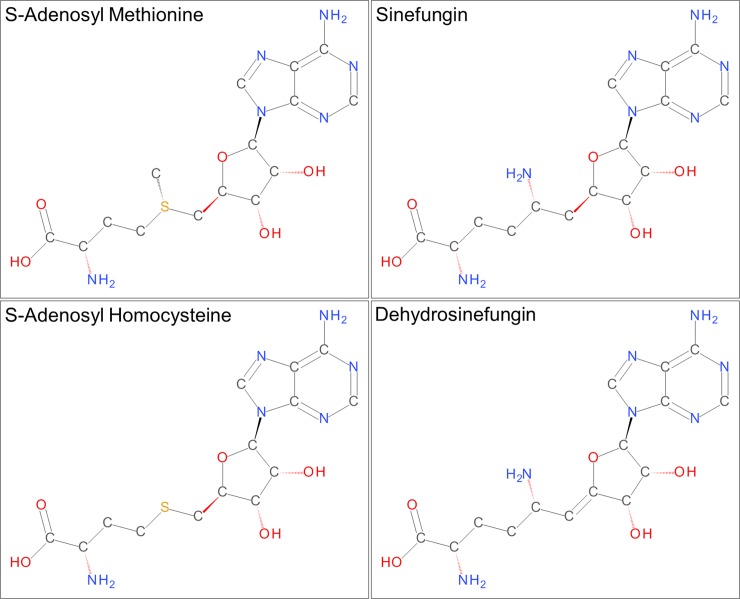
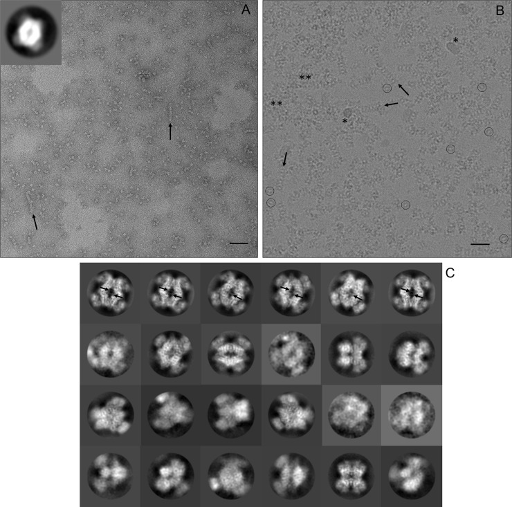
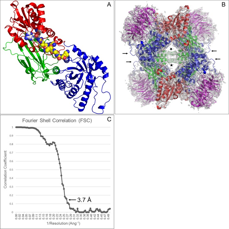
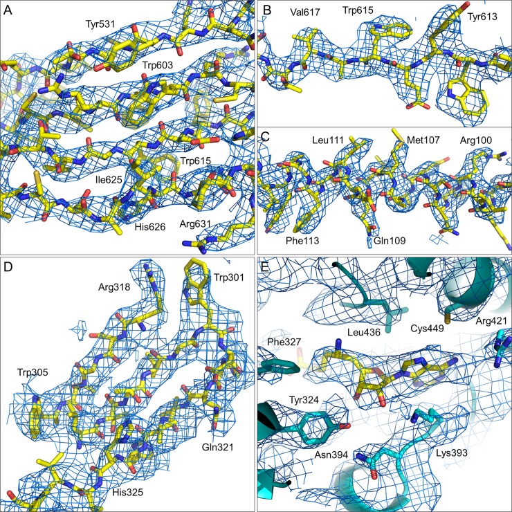
Protein arginine methyl transferase 5 (PRMT5) is a signaling protein and histone modifying enzyme that is important in many cellular processes, including regulation of eukaryotic gene transcription. Reported here is a 3.7 Å structure of PRMT5, solved in complex with regulatory binding subunit MEP50 (methylosome associated protein 50, WDR77, p44), by single particle (SP) cryo-Electron Microscopy (cryo-EM) using micrographs of particles that are visibly crowded and aggregated. Despite suboptimal micrograph appearance, this cryo-EM structure is in good agreement with previously reported crystal structures of the complex, which revealed a 450 kDa hetero-octameric assembly having internal D2 symmetry. The catalytic PRMT5 subunits form a core tetramer and the MEP50 subunits are arranged peripherally in complex with the PRMT5 N-terminal domain. The cryo-EM reconstruction shows good side chain definition and shows a well-resolved peak for a bound dehydrosinefungin inhibitor molecule. These results demonstrate the applicability of cryo-EM in determining structures of human protein complexes of biomedical significance and suggests cryo-EM could be further utilized to understand PRMT5 interactions with other biologically important binding proteins and ligands.
Conflict of interest statement
Figures
Similar articles
-
Cryo-EM structure-based selection of computed ligand poses enables design of MTA-synergic PRMT5 inhibitors of better potency.Commun Biol. 2022 Oct 3;5(1):1054. doi: 10.1038/s42003-022-03991-9. Commun Biol. 2022. PMID: 36192627 Free PMC article.
-
Crystal structure of the human PRMT5:MEP50 complex.Proc Natl Acad Sci U S A. 2012 Oct 30;109(44):17960-5. doi: 10.1073/pnas.1209814109. Epub 2012 Oct 15. Proc Natl Acad Sci U S A. 2012. PMID: 23071334 Free PMC article.
-
Histone H2A and H4 N-terminal tails are positioned by the MEP50 WD repeat protein for efficient methylation by the PRMT5 arginine methyltransferase.J Biol Chem. 2015 Apr 10;290(15):9674-89. doi: 10.1074/jbc.M115.636894. Epub 2015 Feb 24. J Biol Chem. 2015. PMID: 25713080 Free PMC article.
-
The Structure and Function of the PRMT5:MEP50 Complex.Subcell Biochem. 2017;83:185-194. doi: 10.1007/978-3-319-46503-6_7. Subcell Biochem. 2017. PMID: 28271477 Review.
-
The PRMT5 arginine methyltransferase: many roles in development, cancer and beyond.Cell Mol Life Sci. 2015 Jun;72(11):2041-59. doi: 10.1007/s00018-015-1847-9. Epub 2015 Feb 7. Cell Mol Life Sci. 2015. PMID: 25662273 Free PMC article. Review.
Cited by
-
The final step of 40S ribosomal subunit maturation is controlled by a dual key lock.Elife. 2021 Apr 28;10:e61254. doi: 10.7554/eLife.61254. Elife. 2021. PMID: 33908345 Free PMC article.
-
Cryo-EM structure-based selection of computed ligand poses enables design of MTA-synergic PRMT5 inhibitors of better potency.Commun Biol. 2022 Oct 3;5(1):1054. doi: 10.1038/s42003-022-03991-9. Commun Biol. 2022. PMID: 36192627 Free PMC article.
-
Protein arginine methyltransferases: insights into the enzyme structure and mechanism at the atomic level.Cell Mol Life Sci. 2019 Aug;76(15):2917-2932. doi: 10.1007/s00018-019-03145-x. Epub 2019 May 23. Cell Mol Life Sci. 2019. PMID: 31123777 Free PMC article. Review.
-
Discovery and Biological Characterization of PRMT5:MEP50 Protein-Protein Interaction Inhibitors.J Med Chem. 2022 Oct 27;65(20):13793-13812. doi: 10.1021/acs.jmedchem.2c01000. Epub 2022 Oct 7. J Med Chem. 2022. PMID: 36206451 Free PMC article.
-
Discovery of a First-in-Class Inhibitor of the PRMT5-Substrate Adaptor Interaction.J Med Chem. 2021 Aug 12;64(15):11148-11168. doi: 10.1021/acs.jmedchem.1c00507. Epub 2021 Aug 3. J Med Chem. 2021. PMID: 34342224 Free PMC article.
References
-
- Di Lorenzo A, Bedford MT. Histone arginine methylation. FEBS Lett. 2011;585(13):2024–31. doi: 10.1016/j.febslet.2010.11.010 - DOI - PMC - PubMed
-
- Friesen WJ, Paushkin S, Wyce A, Massenet S, Pesiridis GS, Van Duyne G, et al. The methylosome, a 20S complex containing JBP1 and pICln, produces dimethylarginine-modified Sm proteins. Mol Cell Biol. 2001;21(24):8289–300. doi: 10.1128/MCB.21.24.8289-8300.2001 - DOI - PMC - PubMed
-
- Burgos ES, Wilczek C, Onikubo T, Bonanno JB, Jansong J, Reimer U, et al. Histone H2A and H4 N-terminal tails are positioned by the MEP50 WD repeat protein for efficient methylation by the PRMT5 arginine methyltransferase. J Biol Chem. 2015;290(15):9674–89. doi: 10.1074/jbc.M115.636894 - DOI - PMC - PubMed
-
- Fabbrizio E, El Messaoudi S, Polanowska J, Paul C, Cook JR, Lee JH, et al. Negative regulation of transcription by the type II arginine methyltransferase PRMT5. EMBO Rep. 2002;3(7):641–5. doi: 10.1093/embo-reports/kvf136 - DOI - PMC - PubMed
-
- Wang L, Pal S, Sif S. Protein arginine methyltransferase 5 suppresses the transcription of the RB family of tumor suppressors in leukemia and lymphoma cells. Mol Cell Biol. 2008;28(20):6262–77. doi: 10.1128/MCB.00923-08 - DOI - PMC - PubMed
Publication types
MeSH terms
Substances
LinkOut - more resources
Full Text Sources
Other Literature Sources
