. 2018 Mar 7;97(5):1023-1031.e7.
doi: 10.1016/j.neuron.2018.01.031.

TREM2 Is a Receptor for β-Amyloid that Mediates Microglial Function

Affiliations

TREM2 Is a Receptor for β-Amyloid that Mediates Microglial Function

Yingjun Zhao et al. Neuron. .

Abstract

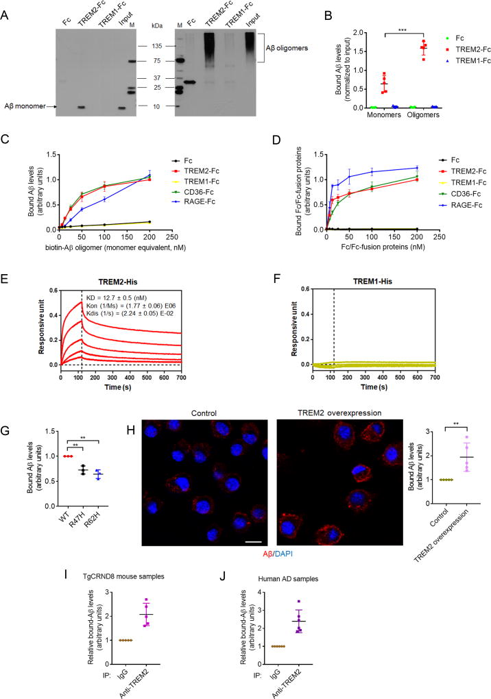
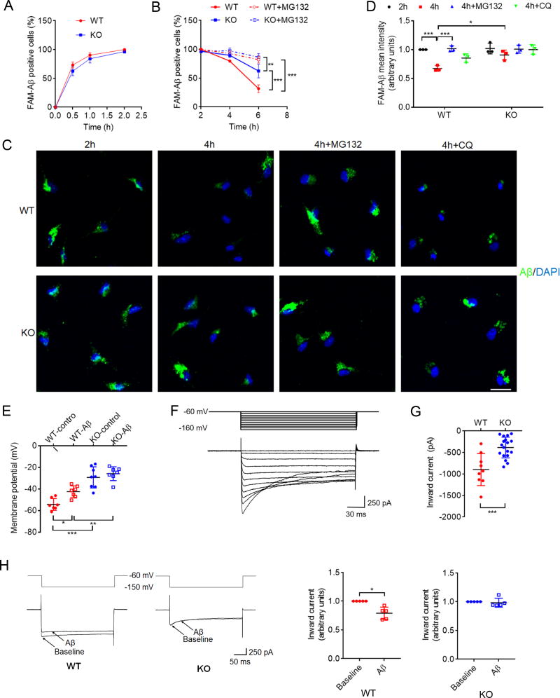
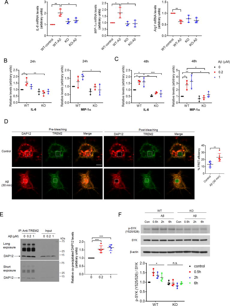
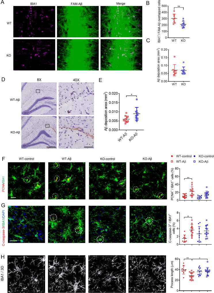
Mutations in triggering receptor expressed on myeloid cells 2 (TREM2) have been linked to increased Alzheimer's disease (AD) risk. Neurobiological functions of TREM2 and its pathophysiological ligands remain elusive. Here we found that TREM2 directly binds to β-amyloid (Aβ) oligomers with nanomolar affinity, whereas AD-associated TREM2 mutations reduce Aβ binding. TREM2 deficiency impairs Aβ degradation in primary microglial culture and mouse brain. Aβ-induced microglial depolarization, K+ inward current induction, cytokine expression and secretion, migration, proliferation, apoptosis, and morphological changes are dependent on TREM2. In addition, TREM2 interaction with its signaling adaptor DAP12 is enhanced by Aβ, regulating downstream phosphorylation of SYK and GSK3β. Our data demonstrate TREM2 as a microglial Aβ receptor transducing physiological and AD-related pathological effects associated with Aβ.

Keywords: Alzheimer’s disease; Aβ; R47H; TREM2; microglia; neuroinflammation.


Conflict of interest statement

DECLARATION OF INTERESTS

The authors declare no competing interests.

Figures

Figure 1
Figure 1. TREM2 binds to Aβ
(A and B) The binding of Aβ1–42 monomers or oligomers with Fc, TREM2-Fc or TREM1-Fc. All values were normalized to input. M: marker. n=5 independent experiments, ***P<.001 by unpaired t-test. (C) Levels of biotin-oAβ1–42 bound to immobilized Fc/Fc-fusion proteins were normalized to maximal Aβ/TREM2-Fc interactions. n = 3 independent experiments. (D) Levels of Fc/Fc-fusion proteins bound to immobilized biotin-oAβ1–42 were normalized to maximal TREM2-Fc/Aβ binding. n = 3 independent experiments. (E and F) Bio-Layer interferometry binding kinetics of TREM2-His (E) or TREM1-His (F) incubated with immobilized biotin-oAβ1–42 (curves correspond to His-tagged proteins with the following concentrations: 222.2nM (top), 74.1nM, 24.7nM, 8.23nM and 2.74nM (bottom); in E and F). (G) Maximal levels of biotin-oAβ1–42 bound to immobilized wild type (WT) TREM2-Fc, R47H or R62H mutants (normalized to WT). n = 3 independent experiments, **P < 0.01, one-way ANOVA followed by Dunnett’s post hoc test. (H) Representative images of biotin-oAβ1–42 (red) bound to BV2 cells overexpressing TREM2 or BV2 control (DAPI, blue; scale bar = 10 µm). n = 5 independent experiments, **P < 0.01, unpaired t-test. (I and J) Levels of Aβ1–42 co-precipitated with TREM2 in lysates from APP transgenic TgCRND8 mice (I) or human AD brains (J) were determined by ELISA. Bound Aβ was normalized to the IgG control from each sample (n = 5 for TgCRND8, n = 6 for human AD). All values represent mean ± s.d. See also Figure S1 and Table S1.
Figure 2
Figure 2. TREM2 deficiency modulates microglia-mediated Aβ degradation, and Aβ-induced depolarization
(A–D) TREM2 deficiency attenuates microglia-mediated Aβ degradation. Cultured microglia from WT or TREM2 KO mice were incubated with 200 nM FAM-Aβ1–42 aggregates at 37°C for as the time indicated in (A), and washed/re-incubated in Aβ-free media with or without MG132 (B–D) or chloroquine (CQ) (C and D). FAM-Aβ in WT or TREM2 KO microglia at different time points was determined by flow cytometry (A and B), or confocal microscopy (C). All values were normalized to WT FAM-Aβ levels 2h after uptake. Quantification of FAM-Aβ fluorescence is shown in (D). Scale bar=20 µm. n= 3 independent measurements, *P < 0.05, **P < 0.01, ***P < 0.001, two-way ANOVA followed by Tukey post hoc test. (E) Resting membrane potential in WT and TREM2 KO microglia with or without 6h exposure to oAβ1–42 (5 µM). WT-control, n = 7; WT-Aβ, n = 8; KO-control, n = 8; KO-Aβ, n = 7. *P < 0.05, **P < 0.01, ***P < 0.001, two-way ANOVA followed by Tukey’s post hoc test. (F) Representative inward currents obtained from primary microglia. (G) TREM2 KO microglia showed significantly reduced peak inward currents (n = 18 cells) when compared to WT (n = 9 cells). ***P < 0.001, unpaired t-test. (H) Aβ1–42 oligomers (10 min) reduce current amplitude in WT but not in TREM2 KO microglia. At −150 mV, current amplitude was significantly reduced in WT but not in TREM2 KO microglia. All values were normalized to baseline for each cell. n = 5 cells for each group, *P < 0.05, by paired t-test. Electrophysiological recordings were taken from 4 independent microglial cultures. All values represent mean ± s.d. See also Figure S2.
Figure 3
Figure 3. TREM2 deficiency alters microglia cytokine expression/secretion and downstream signaling in response to Aβ
(A) IL-6, MIP-1α, or Arg1 expression was determined by qRT-PCR in WT or TREM2 KO microglia in the presence or absence of 1 µM Aβ. n = 3 independent experiments, *P < 0.05, **P < 0.01, by two-way ANOVA followed by Tukey’s post hoc test. (B and C) Cytokines levels in conditioned media from WT or TREM2 KO microglial cultures treated with oAβ1–42 for 24h (B) or 48h (C) were quantified by ELISA. All values were normalized to WT-Aβ (0 µM) levels for each time point. n = 3 independent experiments. *P < 0.05, **P < 0.01, ***P < 0.001, two-way ANOVA followed by Tukey’s post hoc test. (D) BV2 cells overexpressing TREM2-GFP and DAP12-mCherry were treated with or without (1 µM) for 30 min, and interactions between TREM2-GFP and DAP12-mCherry were determined by FRET analysis. White rectangles indicate bleached regions. Scale bar = 10 µm. n = 6 cells per group. **P < 0.01, unpaired t-test. (E) Levels of endogenous DAP12 co-precipitated with TREM2 in primary microglia treated with oAβ1–42 were determined by Western-blot. n = 5 independent experiments. ***P < 0.001, one-way ANOVA followed by Dunnett’s post hoc test. (F) Levels of tyrosine-phosphorylated (Y525/526) and total SYK in WT or TREM2 KO microglia treated with oAβ1–42 (1 µM) were determined by Western blot. n=3 independent experiments. *P<0.05; n.s., no significance; one-way ANOVA. All values represent mean ± s.d. See also Figure S3.
Figure 4
Figure 4. Microglial migration, Aβ degradation and downstream effects on microglial proliferation and apoptosis are altered in TREM2 KO mice
(A–C) Analysis of WT or TREM2 KO brain at 16 hours after FAM-Aβ injection. n = 8 for WT, n = 9 for KO mice. (A) Representative images of Aβ injected regions. Scale bar = 20 µm. (B)Quantified average values for total Aβ/IBA1 positive (IBA1+) cells. **P < 0.01 by nonparametric t-test. (C) Quantified Aβ deposition area. (D and E) Aβ staining (MOAB-2) in WT or TREM2 KO mouse hippocampus 3 days after oAβ1–42 injection (scale bar = 300 µm for 8X magnification, scale bar = 60 µm for 40× magnification in D). n = 9 for WT, n = 8 for KO mice, *P < 0.05 by nonparametric t-test. (F and G) Immunostaining of PCNA (red) (F), cleaved-caspase-3 (C-caspase-3, red) (G), IBA1 microglial markers (green) and DAPI (blue) in the hippocampus of WT or TREM2 KO mice with control vehicle or oAβ injection as indicated. PCNA or c-caspase-3 positive microglial cells are indicated by white circles. Adjacent graphs represent quantified overlapping IBA1/PCNA (F) or IBA1/C-caspase-3 (G) stained cells from 8–12 mice per group. Scale bar = 20 µm. *P < 0.05, **P < 0.01, two-way ANOVA followed by Tukey’s post hoc test. (H) Reconstructed 3D microscopy images of IBA1-stained (white) microglia. Average process length per microglia was quantified and shown in the adjacent graph. n = 9–12 mice/group. Scale bar = 10 µm ***P < 0.001, two-way ANOVA followed by Tukey’s post hoc test. All graphs represent mean ± s.d. See also Figure S4.

Comment in

References

    1. Askew K, Li K, Olmos-Alonso A, Garcia-Moreno F, Liang Y, Richardson P, Tipton T, Chapman MA, Riecken K, Beccari S, et al. Coupled Proliferation and Apoptosis Maintain the Rapid Turnover of Microglia in the Adult Brain. Cell Rep. 2017;18:391–405. - PMC - PubMed
    1. Atagi Y, Liu CC, Painter MM, Chen XF, Verbeeck C, Zheng H, Li X, Rademakers R, Kang SS, Xu H, et al. Apolipoprotein E Is a Ligand for Triggering Receptor Expressed on Myeloid Cells 2 (TREM2) J Biol Chem. 2015;290:26043–26050. - PMC - PubMed
    1. Boucsein C, Kettenmann H, Nolte C. Electrophysiological properties of microglial cells in normal and pathologic rat brain slices. Eur J Neurosci. 2000;12:2049–2058. - PubMed
    1. Cavanaugh SE, Pippin JJ, Barnard ND. Animal models of Alzheimer disease: historical pitfalls and a path forward. ALTEX. 2014;31:279–302. - PubMed
    1. Chen J, Carey K, Godowski PJ. Identification of Gas6 as a ligand for Mer, a neural cell adhesion molecule related receptor tyrosine kinase implicated in cellular transformation. Oncogene. 1997;14:2033–2039. - PubMed

Publication types

MeSH terms