

Subacute intranasal administration of tissue plasminogen activator improves stroke recovery by inducing axonal remodeling in mice
- PMID: 29518364
- PMCID: PMC5916497
- DOI: 10.1016/j.expneurol.2018.03.001
Subacute intranasal administration of tissue plasminogen activator improves stroke recovery by inducing axonal remodeling in mice
Abstract
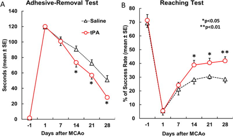
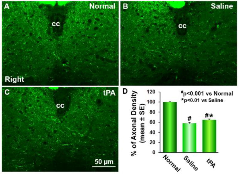
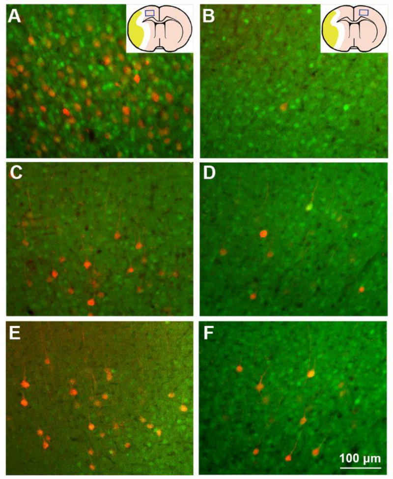
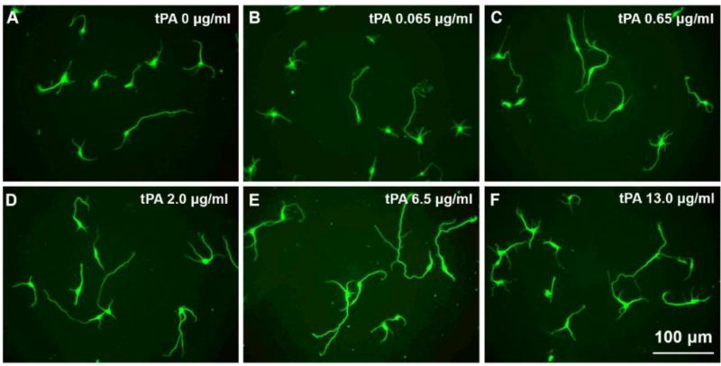
In addition to thrombolysis, tissue plasminogen activator (tPA) can evoke neurorestorative processes. We therefore investigated the therapeutic effect of subacute intranasal administration of tPA post stroke on neurological recovery and on corticospinal innervation in mice. A transgenic mouse line, in which the pyramidal neurons and corticospinal tract (CST) axons are specifically labeled by yellow fluorescent protein (YFP) was employed. Adult CST-YFP mice were subjected to right unilateral middle cerebral artery occlusion (MCAo), and were randomly divided into groups treated with saline or tPA intranasally in the subacute phase. Pseudorabies virus (PRV)-614-monomeric red fluorescent protein (RFP) was injected into the left forelimb. The cervical spinal cord and brain were processed for fluorescent microscopy to detect YFP and RFP labeling. Primary embryonic neurons were cultured with tPA at different concentrations. Neurite length and branch numbers were then measured. In vivo, subacute tPA treatment significantly enhanced functional recovery (p < 0.05), and increased CST density in the denervated gray matter, and in the numbers of PRV-labeled neurons in bilateral cortices. The behavioral performance was significantly correlated with axonal density in the denervated spinal cord. In vitro, both neurite length and branch numbers significantly increased with concentration of tPA (p < 0.05). Our results demonstrate that tPA dose-dependently increases neurite outgrowth and branching of cultured cortical neurons. Subacute intranasal administration of tPA may provide enhance neurological recovery after stroke by promoting CST axonal remodeling.
Keywords: Axonal remodeling; Functional recovery; Middle cerebral artery occlusion; Stroke; Tissue plasminogen activator.
Copyright © 2018 Elsevier Inc. All rights reserved.
Figures
Similar articles
-
Axonal remodeling of the corticospinal tract during neurological recovery after stroke.Neural Regen Res. 2021 May;16(5):939-943. doi: 10.4103/1673-5374.297060. Neural Regen Res. 2021. PMID: 33229733 Free PMC article. Review.
-
Subacute intranasal administration of tissue plasminogen activator increases functional recovery and axonal remodeling after stroke in rats.Neurobiol Dis. 2012 Feb;45(2):804-9. doi: 10.1016/j.nbd.2011.11.004. Epub 2011 Nov 15. Neurobiol Dis. 2012. PMID: 22115941 Free PMC article.
-
Targeted tPA overexpression in denervated spinal motor neurons promotes stroke recovery in mice.J Cereb Blood Flow Metab. 2021 Jan;41(1):92-104. doi: 10.1177/0271678X20901686. Epub 2020 Jan 27. J Cereb Blood Flow Metab. 2021. PMID: 31987011 Free PMC article.
-
Plasminogen deficiency causes reduced corticospinal axonal plasticity and functional recovery after stroke in mice.PLoS One. 2014 Apr 14;9(4):e94505. doi: 10.1371/journal.pone.0094505. eCollection 2014. PLoS One. 2014. PMID: 24732409 Free PMC article.
-
Corticospinal tract: a new hope for the treatment of post-stroke spasticity.Acta Neurol Belg. 2024 Feb;124(1):25-36. doi: 10.1007/s13760-023-02377-w. Epub 2023 Sep 13. Acta Neurol Belg. 2024. PMID: 37704780 Free PMC article. Review.
Cited by
-
Small extracellular vesicles derived from cerebral endothelial cells with elevated microRNA 27a promote ischemic stroke recovery.Neural Regen Res. 2025 Jan 1;20(1):224-233. doi: 10.4103/NRR.NRR-D-22-01292. Epub 2024 Mar 1. Neural Regen Res. 2025. PMID: 38767487 Free PMC article.
-
Sequential Transcriptome Changes in the Penumbra after Ischemic Stroke.Int J Mol Sci. 2019 Dec 16;20(24):6349. doi: 10.3390/ijms20246349. Int J Mol Sci. 2019. PMID: 31888302 Free PMC article.
-
Plasminogen deficiency causes reduced angiogenesis and behavioral recovery after stroke in mice.J Cereb Blood Flow Metab. 2021 Oct;41(10):2583-2592. doi: 10.1177/0271678X211007958. Epub 2021 Apr 14. J Cereb Blood Flow Metab. 2021. PMID: 33853408 Free PMC article.
-
Axonal remodeling of the corticospinal tract during neurological recovery after stroke.Neural Regen Res. 2021 May;16(5):939-943. doi: 10.4103/1673-5374.297060. Neural Regen Res. 2021. PMID: 33229733 Free PMC article. Review.
-
Activation of adult endogenous neurogenesis by a hyaluronic acid collagen gel containing basic fibroblast growth factor promotes remodeling and functional recovery of the injured cerebral cortex.Neural Regen Res. 2025 Oct 1;20(10):2923-2937. doi: 10.4103/NRR.NRR-D-23-01706. Epub 2024 Jun 3. Neural Regen Res. 2025. PMID: 39610105 Free PMC article.
References
-
- Bareyre FM, Kerschensteiner M, Misgeld T, Sanes JR. Transgenic labeling of the corticospinal tract for monitoring axonal responses to spinal cord injury. Nat Med. 2005;11:1355–1360. - PubMed
-
- Carmichael ST. Cellular and molecular mechanisms of neural repair after stroke: making waves. Ann Neurol. 2006;59:735–742. - PubMed
-
- Chen J, Li Y, Wang L, Zhang Z, Lu D, Lu M, Chopp M. Therapeutic benefit of intravenous administration of bone marrow stromal cells after cerebral ischemia in rats. Stroke. 2001;32:1005–1011. - PubMed
Publication types
MeSH terms
Substances
Grants and funding
LinkOut - more resources
Full Text Sources
Other Literature Sources
Medical
