

Hypothalamic redox balance and leptin signaling - Emerging role of selenoproteins
- PMID: 29518483
- PMCID: PMC6123311
- DOI: 10.1016/j.freeradbiomed.2018.02.038
Hypothalamic redox balance and leptin signaling - Emerging role of selenoproteins
Abstract
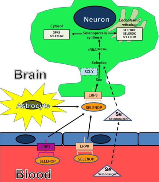
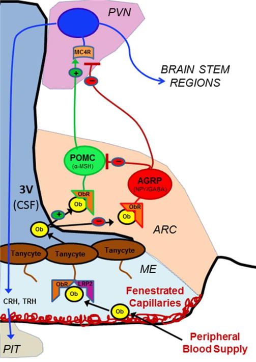
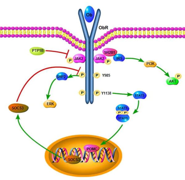
The hypothalamus is the central neural site governing food intake and energy expenditure. During the past 25 years, understanding of the hypothalamic cell types, hormones, and circuitry involved in the regulation of energy metabolism has dramatically increased. It is now well established that the adipocyte-derived hormone, leptin, acts upon two distinct groups of hypothalamic neurons that comprise opposing arms of the central melanocortin system. These two cell populations are anorexigenic neurons expressing proopiomelanocortin (POMC) and orexigenic neurons that express agouti-related peptide (AGRP). Several important studies have demonstrated that reactive oxygen species and endoplasmic reticulum stress significantly impact these hypothalamic neuronal populations that regulate global energy metabolism. Reactive oxygen species and redox homeostasis are influenced by selenoproteins, an essential class of proteins that incorporate selenium co-translationally in the form of the 21st amino acid, selenocysteine. Levels of these proteins are regulated by dietary selenium intake and they are widely expressed in the brain. Of additional relevance, selenium supplementation has been linked to metabolic alterations in both animal and human studies. Recent evidence also indicates that hypothalamic selenoproteins are significant modulators of energy metabolism in both neurons and tanycytes, a population of glial-like cells lining the floor of the 3rd ventricle within the hypothalamus. This review article will summarize current understanding of the regulatory influence of redox status on hypothalamic nutrient sensing and highlight recent work revealing the importance of selenoproteins in the hypothalamus.
Keywords: Endoplasmic reticulum stress; Energy metabolism; Hypothalamus; Leptin; Obesity; Selenoprotein.
Copyright © 2018 Elsevier Inc. All rights reserved.
Figures
Similar articles
-
AMPK is essential for energy homeostasis regulation and glucose sensing by POMC and AgRP neurons.J Clin Invest. 2007 Aug;117(8):2325-36. doi: 10.1172/JCI31516. J Clin Invest. 2007. PMID: 17671657 Free PMC article.
-
Selenoprotein M Promotes Hypothalamic Leptin Signaling and Thioredoxin Antioxidant Activity.Antioxid Redox Signal. 2021 Oct 1;35(10):775-787. doi: 10.1089/ars.2018.7594. Epub 2019 Mar 6. Antioxid Redox Signal. 2021. PMID: 30648404 Free PMC article.
-
Female Mice with Selenocysteine tRNA Deletion in Agrp Neurons Maintain Leptin Sensitivity and Resist Weight Gain While on a High-Fat Diet.Int J Mol Sci. 2021 Oct 12;22(20):11010. doi: 10.3390/ijms222011010. Int J Mol Sci. 2021. PMID: 34681674 Free PMC article.
-
Hypothalamic carnitine metabolism integrates nutrient and hormonal feedback to regulate energy homeostasis.Mol Cell Endocrinol. 2015 Dec 15;418 Pt 1:9-16. doi: 10.1016/j.mce.2015.08.002. Epub 2015 Aug 8. Mol Cell Endocrinol. 2015. PMID: 26261054 Review.
-
Leptin signaling, adiposity, and energy balance.Ann N Y Acad Sci. 2002 Jun;967:379-88. doi: 10.1111/j.1749-6632.2002.tb04293.x. Ann N Y Acad Sci. 2002. PMID: 12079865 Review.
Cited by
-
Prolonged maternal exposure to glucocorticoids alters selenoprotein expression in the developing brain.Front Mol Neurosci. 2023 Mar 24;16:1115993. doi: 10.3389/fnmol.2023.1115993. eCollection 2023. Front Mol Neurosci. 2023. PMID: 37033382 Free PMC article.
-
Biogenic Selenium Nanoparticles in Biomedical Sciences: Properties, Current Trends, Novel Opportunities and Emerging Challenges in Theranostic Nanomedicine.Nanomaterials (Basel). 2023 Jan 19;13(3):424. doi: 10.3390/nano13030424. Nanomaterials (Basel). 2023. PMID: 36770385 Free PMC article. Review.
-
Selenium and Selenoproteins in Adipose Tissue Physiology and Obesity.Biomolecules. 2020 Apr 24;10(4):658. doi: 10.3390/biom10040658. Biomolecules. 2020. PMID: 32344656 Free PMC article. Review.
-
Serum Microelements in Early Pregnancy and their Risk of Large-for-Gestational Age Birth Weight.Nutrients. 2020 Mar 24;12(3):866. doi: 10.3390/nu12030866. Nutrients. 2020. PMID: 32213887 Free PMC article.
-
Fetal Programming Is Deeply Related to Maternal Selenium Status and Oxidative Balance; Experimental Offspring Health Repercussions.Nutrients. 2021 Jun 18;13(6):2085. doi: 10.3390/nu13062085. Nutrients. 2021. PMID: 34207090 Free PMC article. Review.
References
-
- Mokdad AH, et al. The spread of the obesity epidemic in the United States, 1991-1998. JAMA. 1999;282:1519–22. - PubMed
-
- Ozcan L, et al. Endoplasmic reticulum stress plays a central role in development of leptin resistance. Cell Metab. 2009;9:35–51. - PubMed
-
- Eletto D, Chevet E, Argon Y, Appenzeller-Herzog C. Redox controls UPR to control redox. J Cell Sci. 2014;127:3649–58. - PubMed
Publication types
MeSH terms
Substances
Grants and funding
LinkOut - more resources
Full Text Sources
Other Literature Sources
Miscellaneous
