. 2018 Mar 8;15(1):73.
doi: 10.1186/s12974-018-1100-1.

The glycoprotein GPNMB attenuates astrocyte inflammatory responses through the CD44 receptor

Affiliations

The glycoprotein GPNMB attenuates astrocyte inflammatory responses through the CD44 receptor

Matthew L Neal et al. J Neuroinflammation. .

Abstract

Background: Neuroinflammation is one of the hallmarks of neurodegenerative diseases, such as Parkinson's disease (PD). Activation of glial cells, including microglia and astrocytes, is a characteristic of the inflammatory response. Glycoprotein non-metastatic melanoma protein B (GPNMB) is a transmembrane glycoprotein that releases a soluble signaling peptide when cleaved by ADAM10 or other extracellular proteases. GPNMB has demonstrated a neuroprotective role in animal models of ALS and ischemia. However, the mechanism of this protection has not been well established. CD44 is a receptor expressed on astrocytes that can bind GPNMB, and CD44 activation has been demonstrated to reduce NFκB activation and subsequent inflammatory responses in macrophages. GPNMB signaling has not been investigated in models of PD or specifically in astrocytes. More recently, genetic studies have linked polymorphisms in GPNMB with risk for PD. Therefore, it is important to understand the role this signaling protein plays in PD.

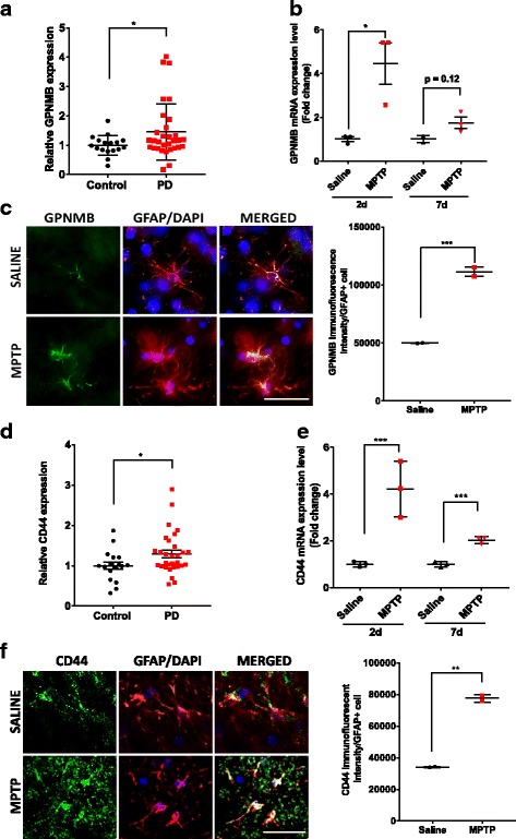
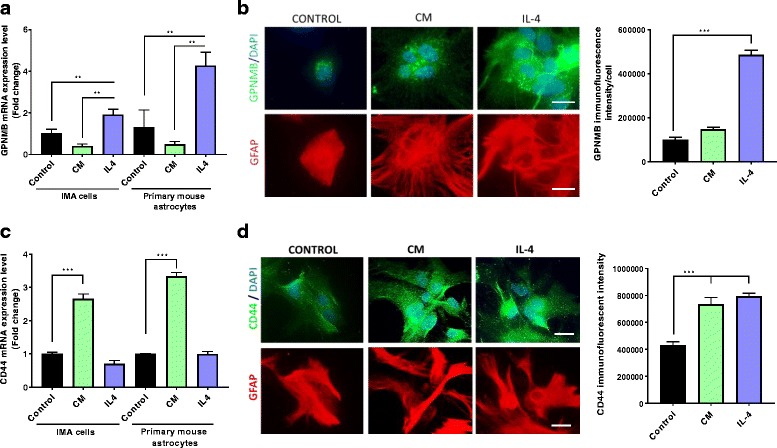
Methods: We used data mining techniques to evaluate mRNA expression of GPNMB and its receptor CD44 in the substantia nigra of PD and control brains. Immunofluorescence and qPCR techniques were used to assess GPNMB and CD44 levels in mice treated with MPTP. In vitro experiments utilized the immortalized mouse astrocyte cell line IMA2.1 and purified primary mouse astrocytes. The effects of recombinant GPNMB on cytokine-induced astrocyte activation was determined by qPCR, immunofluorescence, and measurement of nitric oxide and reactive oxygen production.

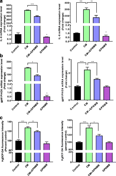
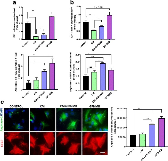
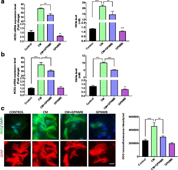
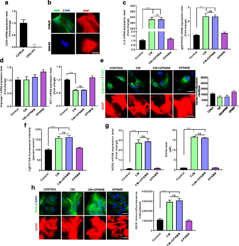
Results: Increased GPNMB and CD44 expression was observed in the substantia nigra of human PD brains and in GFAP-positive astrocytes in an animal model of PD. GPNMB treatment attenuated cytokine-induced levels of inducible nitric oxide synthase, nitric oxide, reactive oxygen species, and the inflammatory cytokine IL-6 in an astrocyte cell line and primary mouse astrocytes. Using primary mouse astrocytes from CD44 knockout mice, we found that the anti-inflammatory effects of GPNMB are CD44-mediated.

Conclusions: These results demonstrate that GPNMB may exert its neuroprotective effect through reducing astrocyte-mediated neuroinflammation in a CD44-dependent manner, providing novel mechanistic insight into the neuroprotective properties of GPNMB.

Keywords: Astrocyte; CD44; GPNMB; Neuroinflammation; Parkinson’s disease.


Conflict of interest statement

Ethics approval and consent to participate

All procedures were performed in accordance with National Institutes of Health guidelines for the care and use of laboratory animals with approval by the Institutional Animal Care and Use Committee of Northeast Ohio Medical University. Gene expression data contained no identifying material and were publicly available.

Consent for publication

Not applicable.

Competing interests

Dr. Safadi has patent and commercialization interests in GPNMB as a therapeutic in bone healing through his company GPN Therapeutics. The remaining authors declare that they have no competing interests.

Publisher’s Note

Springer Nature remains neutral with regard to jurisdictional claims in published maps and institutional affiliations.

Figures

Fig. 1
Fig. 1
Increased GPNMB and CD44 levels in PD patients and an acute MPTP mouse model. a Gene expression of GPNMB is increased in the substantia nigra of PD patients (n = 22 males and n = 9 females) compared to age-matched controls (n = 10 males and n = 7 females). GEO numbers were normalized to 1 to generate relative expression values and allow comparison between datasets. b GPNMB gene expression in the mouse striatum by qPCR following an acute injection of MPTP in mice and sacrificed at 2 and 7 days post injection. c Representative images of immunofluorescent confocal microscopy of GPNMB (green), GFAP (red), and DAPI nuclear stain (blue) at × 40 magnification following an acute injection of saline or MPTP, scale bar and 50 μm. d Gene expression of CD44 in human PD patients examined by microarray on the GEO demonstrating a significant increase in CD44 gene expression in the SN of PD patients. e–f CD44 gene expression and protein levels in the mouse striatum by e qPCR and immunofluorescent confocal microscopy of CD44 (green), GFAP (red), and DAPI nuclear stain (blue) at f × 40 magnification following an acute injection of Saline or MPTP, scale bar and 50 μm. Asterisks denote statistically significant differences between PD and age-matched controls or MPTP and saline controls (*p < 0.05, **p < 0.01, and ***p < 0.001)
Fig. 2
Fig. 2
Increased GPNMB and CD44 in cultured astrocytes exposed to different stimuli. a–b GPNMB gene expression (a) in cultured IMA2.1 cells and PMAs by qPCR, and protein levels (b) by immunofluorescent microscopy in PMAs (GPNMB in green, DAPI in blue, and GFAP in red). c CD44 gene expression in IMA2.1 cells and PMAs treated with or without the inflammatory cytokine mix (CM) or recombinant IL-4. d Immunofluorescence images for CD44 (green) protein levels in GFAP-positive (red) primary mouse astrocytes following CM or IL-4 treatment (scale bar 20 μm). Asterisks denote statistically significant differences between CM or IL-4 treatment and control (**p < 0.01 and ***p < 0.001)
Fig. 3
Fig. 3
GPNMB reduces inflammatory mediators in cultured astrocytes. a–b Gene expression of the inflammatory factors IL-6 (a) and gp91phox (b) in IMA cells (left panel) and PMAs (right panel). c H2DCF-DA fluorescence intensity measuring intracellular ROS generation in IMA (left panel) and PMA (right panel) cells. Asterisks denote statistically significant differences between the different treatment groups or compared to the control (*p < 0.05, **p < 0.01, and ***p < 0.001)
Fig. 4
Fig. 4
rGPNMB induces anti-inflammatory factors IGF-1 and arginase-1 in cultured astrocytes. a–b Gene expression of the anti-inflammatory factors IGF-1 (top panel) and arginase-1 (bottom panel) in IMA cells (a) and PMAs (b). c Representative immunofluorescent images and quantification for arginase-1 in PMAs treated with CM alone, GPNMB alone or co-treated with CM and GPNMB (scale bar 20 μm). Asterisks denote statistically significant differences between the different treatment groups or compared to the control (*p < 0.05, **p < 0.01, and ***p < 0.001)
Fig. 5
Fig. 5
GPNMB attenuated cytokine-induced reactive nitrogen species production. a–b Gene expression of NOS2 (left panel) and cell culture supernatant nitrite levels (right panel) from IMA cells (a) and PMAs (b) treated with either CM, GPNMB, or co-treated with both. c Representative immunofluorescent images and quantification for iNOS protein levels in PMAs treated with CM, GPNMB, or co-treated with both (scale bar 20 μm). Asterisks denote statistically significant differences between the different treatment groups or compared to the control (*p < 0.05, **p < 0.01, and ***p < 0.001)
Fig. 6
Fig. 6
CD44 KO attenuates the GPNMB-induced reduction of cultured astrocyte inflammatory response. ab Isolated CD44 KO PMAs do not have CD44 gene expression (a) or any visible CD44 protein levels (b) by immunofluorescent imaging (scale bar 20 μm). c Gene expression of the inflammatory factors IL-6 (left panel) and gp91phox (right panel) by qPCR in CD44 KO PMAs. d Gene expression of the anti-inflammatory factors arginase-1 (left panel) and IGF-1 (right panel) by qPCR in CD44 KO PMAs. e Representative immunofluorescent images for arginase-1 in CD44 KO PMAs treated with CM, rGPNMB or co-treated with both (scale bar 20 μm). f H2DCF-DA fluorescence intensity measuring intracellular ROS generation in CD44 KO PMAs. gh rGPNMB treatment failed to attenuate CM-induced NOS2 gene expression (g, left panel), supernatant nitrite levels (g, right panel), and iNOS protein levels (h) in CD44 KO PMAs. Asterisks denote statistically significant differences between the different treatment groups or compared to the control (*p < 0.05 and ***p < 0.001, not statistically significant groups are represented with ns)

References

    1. Tanner CM, Goldman SM. Epidemiology of Parkinson’s disease. Neurol Clin. 1996;14:317–335. doi: 10.1016/S0733-8619(05)70259-0. - DOI - PMC - PubMed
    1. Nagatsu T, Sawada M. Cellular and molecular mechanisms of Parkinson’s disease: neurotoxins, causative genes, and inflammatory cytokines. Cell Mol Neurobiol. 2006;26:781–802. doi: 10.1007/s10571-006-9061-9. - DOI - PMC - PubMed
    1. Yasuda Y, Shimoda T, Uno K, Tateishi N, Furuya S, Yagi K, Suzuki K, Fujita S. The effects of MPTP on the activation of microglia/astrocytes and cytokine/chemokine levels in different mice strains. J Neuroimmunol. 2008;204:43–51. doi: 10.1016/j.jneuroim.2008.08.003. - DOI - PubMed
    1. Teismann P, Tieu K, Cohen O, Choi DK, Wu DC, Marks D, Vila M, Jackson-Lewis V, Przedborski S. Pathogenic role of glial cells in Parkinson’s disease. Mov Disord. 2003;18:121–129. doi: 10.1002/mds.10332. - DOI - PubMed
    1. Qian L, Flood PM. Microglial cells and Parkinson’s disease. Immunol Res. 2008;41:155–164. doi: 10.1007/s12026-008-8018-0. - DOI - PubMed

MeSH terms