. 2018 Apr;120(3):282-291.
doi: 10.1016/j.acthis.2018.02.010. Epub 2018 Mar 5.

A novel surgical technique for a rat subcutaneous implantation of a tissue engineered scaffold

Affiliations

A novel surgical technique for a rat subcutaneous implantation of a tissue engineered scaffold

Reza Khorramirouz et al. Acta Histochem. 2018 Apr.

Abstract

Objectives: Subcutaneous implantations in small animal models are currently required for preclinical studies of acellular tissue to evaluate biocompatibility, including host recellularization and immunogenic reactivity.

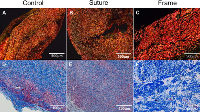
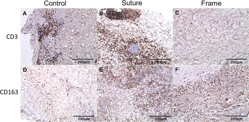
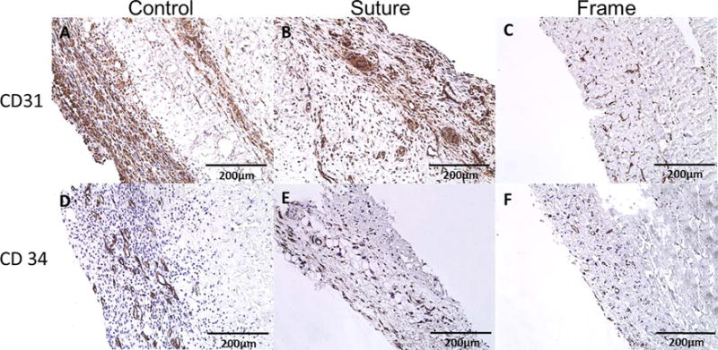
Methods: Three rat subcutaneous implantation methods were evaluated in six Sprague Dawley rats. An acellular xenograft made from porcine pericardium was used as the tissue-scaffold. Three implantation methods were performed; 1) Suture method is where a tissue-scaffold was implanted by suturing its border to the external oblique muscle, 2) Control method is where a tissue-scaffold was implanted without any suturing or support, 3) Frame method is where a tissue-scaffold was attached to a circular frame composed of polycaprolactone (PCL) biomaterial and placed subcutaneously. After 1 and 4 weeks, tissue-scaffolds were explanted and evaluated by hematoxylin and eosin (H&E), Masson's trichrome,Picrosirius Red, transmission electron microscopy (TEM), immunohistochemistry, and mechanical testing.

Results: Macroscopically, tissue-scaffold degradation with the suture method and tissue-scaffold folding with the control method were observed after 4 weeks. In comparison, the frame method demonstrated intact tissue scaffolds after 4 weeks. H&E staining showed progressive cell repopulation over the course of the experiment in all groups with acute and chronic inflammation observed in suture and control methods throughout the duration of the study. Immunohistochemistry quantification of CD3, CD 31, CD 34, CD 163, and αSMA showed a statistically significant differences between the suture, control and frame methods (P < 0.05) at both time points. The average tensile strength was 4.03 ± 0.49, 7.45 ± 0.49 and 5.72 ± 1.34 (MPa) after 1 week and 0.55 ± 0.26, 0.12 ± 0.03 and 0.41 ± 0.32 (MPa) after 4 weeks in the suture, control, and frame methods; respectively. TEM analysis showed an increase in inflammatory cells in both suture and control methods following implantation.

Conclusion: Rat subcutaneous implantation with the frame method was performed with success and ease. The surgical approach used for the frame technique was found to be the best methodology for in vivo evaluation of tissue engineered acellular scaffolds, where the frame method did not compromise mechanical strength, but it reduced inflammation significantly.

Keywords: Acellular xenograft; Extracellular matrix; Histology; Inflammation; Mechanical behavior; Subcutaneous implantation.


Conflict of interest statement

Conflict of interest

There are no financial interests or relationships with industry associated with this paper.

Figures

Fig. 1
Fig. 1
In vivo implantation of pericardial scaffolds including methods utilized. Decellularized and sterilized porcine pericardial scaffolds were cut into 20 mm × 20 mm segments and placed subcutaneously A) without suturing or fixation that showed scaffold folding and shrinkage after 4 weeks denoted with black arrow. B) over the muscle sutured at four corners with Polypropylene 5-0 that showed integration into the host within 1-week post-explant and degradation after 4 weeks, and C) fixed to a PCL rim frame by Polypropylene 5-0 under aseptic conditions, placed between skin and fascia, and left in position that showed no gross changes after 4 weeks.
Fig. 2
Fig. 2
Hematoxylin & Eosin and DAPI stains showing cellularity and DNA content of pericardial scaffolds at 4 weeks. (A–C) H&E at 20 × showed increased cellular infiltration with indication of acute and chronic inflammatory cells in control and suture methods. Frame method showed progressive recellularization overtime. (D–F) DAPI at 4× magnification showed progressive recellularization for all methods, but increased infiltration in both control and suture methods.
Fig. 3
Fig. 3
Collagen deposition. (A–C) Picrosirius staining showed decreased collagen content at 4 weeks. Comparison of collagen content between groups showed increased content in frame methods compared to suture and control methods. (D–F) Masson trichrome staining showed increased inflammatory cell in control and suture groups that invaded the collagenous structure. The frame method showed no evidence of inflammatory cells and intact collagen structure in frame method.
Fig. 4
Fig. 4
Transmission Electron Microscopy at Weeks 1 and 4. (A–F) TEM analysis showed presence of inflammatory cells composed of neutrophil and macrophages (white arrows). (A,D) Control method contained increased neutrophils post implantation. (E) Suture method showed presence of inflammatory cells at 4 weeks but not at 1 week, and (C,F)In the frame method, there were no inflammatory cells present.
Fig. 5
Fig. 5
Uniaxial mechanical testing of 3-methods at all time points. The tensile stress test showed significant decrease from 1 to 4 weeks in all methods. The control method and frame method has more tensile stress in 1 weeks but the control methods tensile stress drop after 4 weeks. At 4 weeks the suture and frame methods has highest tensile test compared to control methods.
Fig. 6
Fig. 6
Immunohistochemistry of inflammation with CD3 and CD163 biomarkers after 1 Week Post-Explantation. The CD 3 staining showed increase inflammatory cells in both the (A) control and (B) suture methods compared to the (C) frame method. Specific staining for macrophage type 2 utilizing CD 163 revealed very weak staining after 1 week in (D) control, (E) suture, and (F) frame methods.
Fig. 7
Fig. 7
Immunohistochemistry of inflammation with CD3 and CD163 biomarkers. (A–B) Positive expression of the CD3 biomarker in both the control and suture methods after 4 weeks showing chronic inflammatory lymphocytes compared to (C) the frame method with no expression of CD3 suggesting no inflammation. CD 163 biomarker, specific for macrophage subtype II, showed a positive expression in (E) suture and (F) frame methods suggestive of increased regenerative capacity over time.
Fig. 8
Fig. 8
Immunohistochemistry of inflammation with CD31 and CD34 biomarkers after 1 Week Post-Explantation. Angiogenesis markers CD 31 and CD 34 showed highly positive staining in the (A,D) control and (B,E) suture methods.
Fig. 9
Fig. 9
Immunohistochemistry of endothelial-like cells with CD31 and CD34 biomarkers at 4 weeks. (A–B) CD31 biomarkers showed a positive expression indicating endothelialization of neovasculature. C) Minimal expression of CD31 in frame method of implantation. (D–F) CD34 biomakers showing expression of angiogenesis in all three methods throughout the study duration.
Fig. 10
Fig. 10
Immunohistochemical staining of interstitial-like cells with Alpha-Smooth Muscle Actin and Vimentin biomarkers. Alpha Smooth muscle actin staining showed highly expressed level after 4 weeks in (A) control and (B) suture methods, which indicate presence of myofibroblast-like interstitial cell infiltration. Vimentin positive cells showed progressive increase 4 weeks in all methods (D–F).
Fig. 11
Fig. 11
Immunohistochemistry of inflammation with Alpha-SMA and Vimentin biomarkers after 1 Week Post-Explantation. Alpha-SMA staining showed that there was moderate positive staining in (A) control and (B) suture methods; however, there was mild positive staining of the (C) frame method tissue. Vimentin staining showed mild positive staining in (D) control, (E) suture, and (F) frame methods at 1 week.

Similar articles

Cited by

References

    1. Şencan A, et al. Testis fixation in prepubertal rats: fibrin glue versus transparenchymal sutures reduces testicular damage. Eur J Pediatr Surg. 2004;14(03):193–197. - PubMed
    1. Ainslie KM, et al. In vitro inflammatory response of nanostructured titania: silicon oxide, and polycaprolactone. J Biomed Mater Res A. 2009;91(3):647–655. - PubMed
    1. Anderson JM. Biological responses to materials. Annu Rev Mater Res. 2001;31(1):81–110.
    1. Andrade MG, Weissman R, Reis SR. Tissue reaction and surface morphology of absorbable sutures after in vivo exposure. J Mater Sci. 2006;17(10):949–961. - PubMed
    1. Ascherman J, Jones V, Knowles S. The histologic effects of retention sutures on wound healing in the rat. Wounds Compend Clin Res Pract. 2005;17(10):271–277.

LinkOut - more resources