

Prenatal Brain MR Imaging: Reference Linear Biometric Centiles between 20 and 24 Gestational Weeks
- PMID: 29519792
- PMCID: PMC7410661
- DOI: 10.3174/ajnr.A5574
Prenatal Brain MR Imaging: Reference Linear Biometric Centiles between 20 and 24 Gestational Weeks
Abstract
Background and purpose: Evaluation of biometry is a fundamental step in prenatal brain MR imaging. While different studies have reported reference centiles for MR imaging biometric data of fetuses in the late second and third trimesters of gestation, no one has reported them in fetuses in the early second trimester. We report centiles of normal MR imaging linear biometric data of a large cohort of fetal brains within 24 weeks of gestation.
Materials and methods: From the data bases of 2 referral centers of fetal medicine, accounting for 3850 examinations, we retrospectively collected 169 prenatal brain MR imaging examinations of singleton pregnancies, between 20 and 24 weeks of gestational age, with normal brain anatomy at MR imaging and normal postnatal neurologic development. To trace the reference centiles, we used the CG-LMS method.
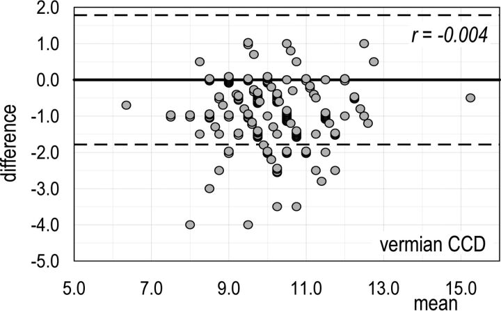
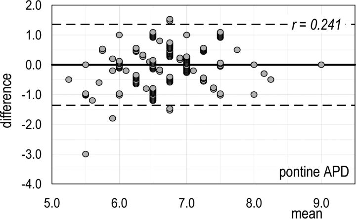
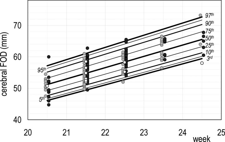
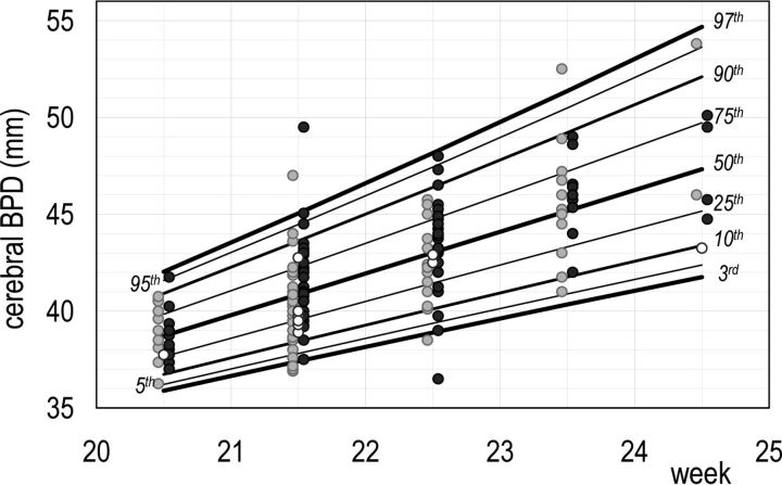
Results: Reference biometric centiles for the developing structures of the cerebrum, cerebellum, brain stem, and theca were obtained. The overall interassessor agreement was adequate for all measurements.
Conclusions: Reference biometric centiles of the brain structures in fetuses between 20 and 24 weeks of gestational age may be a reliable tool in assessing fetal brain development.
© 2018 by American Journal of Neuroradiology.
Figures
Similar articles
-
Reference biometry of foetal brain by prenatal MRI and the distribution of measurements in foetuses with ventricular septal defect.Ann Med. 2021 Dec;53(1):1428-1437. doi: 10.1080/07853890.2021.1969590. Ann Med. 2021. PMID: 34414830 Free PMC article.
-
Development of the Fetal Vermis: New Biometry Reference Data and Comparison of 3 Diagnostic Modalities-3D Ultrasound, 2D Ultrasound, and MR Imaging.AJNR Am J Neuroradiol. 2016 Jul;37(7):1359-66. doi: 10.3174/ajnr.A4725. Epub 2016 Mar 31. AJNR Am J Neuroradiol. 2016. PMID: 27032974 Free PMC article.
-
Prenatal magnetic resonance imaging: brain normal linear biometric values below 24 gestational weeks.Neuroradiology. 2008 Oct;50(10):877-83. doi: 10.1007/s00234-008-0421-7. Epub 2008 Jun 19. Neuroradiology. 2008. PMID: 18563404
-
Fetal growth reference ranges in twin pregnancy: analysis of the Southwest Thames Obstetric Research Collaborative (STORK) multiple pregnancy cohort.Ultrasound Obstet Gynecol. 2015 Mar;45(3):301-7. doi: 10.1002/uog.14640. Epub 2014 Aug 25. Ultrasound Obstet Gynecol. 2015. PMID: 25052857
-
Association of gestational age with MRI-based biometrics of brain development in fetuses.BMC Med Imaging. 2020 Nov 25;20(1):125. doi: 10.1186/s12880-020-00525-9. BMC Med Imaging. 2020. PMID: 33238909 Free PMC article.
Cited by
-
Brain fetal neuroradiology: a beginner's guide.Transl Pediatr. 2021 Apr;10(4):1065-1077. doi: 10.21037/tp-20-293. Transl Pediatr. 2021. PMID: 34012856 Free PMC article. Review.
-
Onset of Chiari type 1 malformation: insights from a small series of intrauterine MR imaging cases.Neuroradiology. 2023 Sep;65(9):1387-1394. doi: 10.1007/s00234-023-03183-0. Epub 2023 Jun 17. Neuroradiology. 2023. PMID: 37329352
-
Reference biometry of foetal brain by prenatal MRI and the distribution of measurements in foetuses with ventricular septal defect.Ann Med. 2021 Dec;53(1):1428-1437. doi: 10.1080/07853890.2021.1969590. Ann Med. 2021. PMID: 34414830 Free PMC article.
-
Evidence of disrupted rhombic lip development in the pathogenesis of Dandy-Walker malformation.Acta Neuropathol. 2021 Oct;142(4):761-776. doi: 10.1007/s00401-021-02355-7. Epub 2021 Aug 4. Acta Neuropathol. 2021. PMID: 34347142 Free PMC article.
-
Comparative neuroimaging of sex differences in human and mouse brain anatomy.Elife. 2024 Mar 15;13:RP92200. doi: 10.7554/eLife.92200. Elife. 2024. PMID: 38488854 Free PMC article.
References
MeSH terms
LinkOut - more resources
Full Text Sources
Other Literature Sources
Medical