

Age-related changes in the peripheral retinal nerve fiber layer thickness
- PMID: 29520130
- PMCID: PMC5833791
- DOI: 10.2147/OPTH.S157429
Age-related changes in the peripheral retinal nerve fiber layer thickness
Abstract
Purpose: This pilot cross-sectional study aimed to determine age-related changes of the retinal nerve fiber layer (RNFL) thickness in retinal periphery by swept-source optical coherence tomography-based analysis.
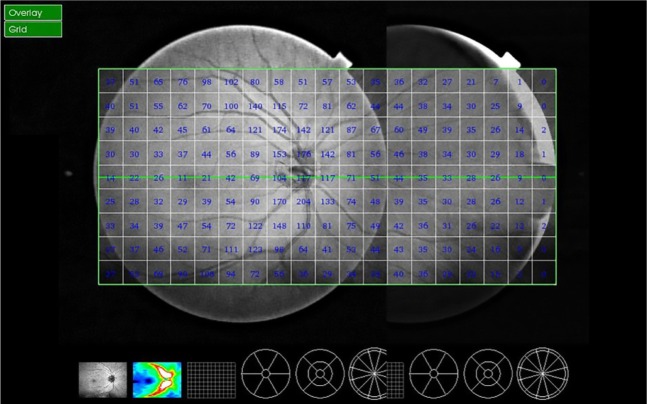
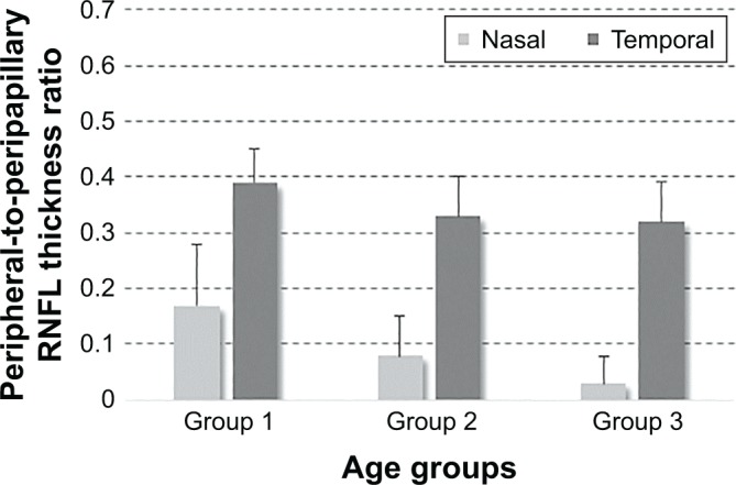
Methods: Forty eyes of 40 healthy subjects were studied in three age groups, group 1 (20-40 years, n=15), group 2 (41-60 years, n=14), and group 3 (≥61 years, n=11). Wide-angle swept-source optical coherence tomography scans, including the optic disc and macula, were montaged with the nasal peripheral optical coherence tomography images acquired with a contralateral gaze. The peripapillary and peripheral RNFL thickness values were obtained for nasal and temporal sides. The ratio of peripheral-to-peripapillary RNFL thickness was also calculated for these sectors.
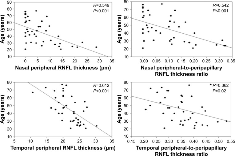
Results: We detected a significantly thinner RNFL in older than younger subjects at a distance of 6 mm from the optic disc on nasal and temporal sides (P<0.001). An age-related reduction in peripheral-to-peripapillary RNFL thickness ratios (P<0.001 and P<0.02 for nasal and temporal sides, respectively) was also detected.
Conclusion: The age-related decline should be taken into consideration when determining the glaucoma-related alterations in peripheral RNFL thickness. Continued analysis in patients with ocular hypertension and glaucoma should help determine whether RNFL in the periphery with lower nerve tissue reserve might be more susceptible to injury, whether injury to the peripheral RNFL might be easier to detect, and/or whether analysis of the peripheral RNFL thickness could improve clinical diagnosis and follow-up of glaucoma.
Keywords: age; glaucoma; retinal nerve fiber layer thickness; swept-source optical coherence tomography.
Conflict of interest statement
Disclosure The authors report no conflicts of interest in this work.
Figures


Similar articles
-
Diagnostic ability of retinal nerve fiber layer imaging by swept-source optical coherence tomography in glaucoma.Am J Ophthalmol. 2015 Jan;159(1):193-201. doi: 10.1016/j.ajo.2014.10.019. Epub 2014 Oct 22. Am J Ophthalmol. 2015. PMID: 25448991 Free PMC article.
-
Peripapillary retinal nerve fiber layer thickness in sickle-cell hemoglobinopathies using spectral-domain optical coherence tomography.Am J Ophthalmol. 2013 Mar;155(3):456-464.e2. doi: 10.1016/j.ajo.2012.09.015. Epub 2012 Dec 4. Am J Ophthalmol. 2013. PMID: 23218697
-
Optical tomography-measured retinal nerve fiber layer thickness in normal latinos.Invest Ophthalmol Vis Sci. 2003 Aug;44(8):3369-73. doi: 10.1167/iovs.02-0975. Invest Ophthalmol Vis Sci. 2003. PMID: 12882783
-
Microvascular and structural alterations in the optic nerve head of advanced primary open-angle glaucoma compared with atrophic non-arteritic anterior ischemic optic neuropathy.Graefes Arch Clin Exp Ophthalmol. 2021 Jul;259(7):1945-1953. doi: 10.1007/s00417-021-05122-2. Epub 2021 Mar 4. Graefes Arch Clin Exp Ophthalmol. 2021. PMID: 33661365
-
Correlation of Peripapillary Choroidal Thickness and Retinal Nerve Fiber Layer Thickness in Normal Subjects and in Patients with Glaucoma.Semin Ophthalmol. 2017;32(5):602-606. doi: 10.3109/08820538.2016.1139736. Epub 2016 Jul 1. Semin Ophthalmol. 2017. PMID: 27367144
Cited by
-
Reversing Aging and Improving Health Span in Glaucoma Patients: The Next Frontier?J Curr Glaucoma Pract. 2024 Jul-Sep;18(3):87-93. doi: 10.5005/jp-journals-10078-1451. Epub 2024 Oct 29. J Curr Glaucoma Pract. 2024. PMID: 39575133 Free PMC article.
-
Investigating possible retinal biomarkers of head trauma in Olympic boxers using optical coherence tomography.Eye Brain. 2018 Dec 14;10:101-110. doi: 10.2147/EB.S183042. eCollection 2018. Eye Brain. 2018. PMID: 30588143 Free PMC article.
-
Selective and Inverse U-Shaped Curve Alteration of the Retinal Nerve in Amyotrophic Lateral Sclerosis: A Potential Mirror of the Disease.Front Aging Neurosci. 2022 Jan 6;13:783431. doi: 10.3389/fnagi.2021.783431. eCollection 2021. Front Aging Neurosci. 2022. PMID: 35069179 Free PMC article.
-
Thicker Retinal Nerve Fiber Layer with Age among Schoolchildren: The Hong Kong Children Eye Study.Diagnostics (Basel). 2022 Feb 15;12(2):500. doi: 10.3390/diagnostics12020500. Diagnostics (Basel). 2022. PMID: 35204590 Free PMC article.
-
An Analysis of Optic Disc Parameters in Patients with Peripheral Retinal Tears Following Acute Posterior Vitreous Detachment: A Cross-Sectional Study.Clin Interv Aging. 2024 Jun 27;19:1153-1162. doi: 10.2147/CIA.S466511. eCollection 2024. Clin Interv Aging. 2024. PMID: 38952872 Free PMC article.
References
-
- Blumenthal EZ, Williams JM, Weinreb RN, Girkin CA, Berry CC, Zangwill LM. Reproducibility of nerve fiber layer thickness measurements by use of optical coherence tomography. Ophthalmology. 2000;107:2278–2282. - PubMed
-
- Hirata M, Tsujikawa A, Matsumoto A, Hangai M, et al. Macular choroidal thickness and volume in normal subjects measured by swept-source optical coherence tomography. Invest Ophthalmol Vis Sci. 2011;52(8):4971–4978. - PubMed
Grants and funding
LinkOut - more resources
Full Text Sources
Other Literature Sources
