

Evaluation of the Subscapularis Tendon Tears on 3T Magnetic Resonance Arthrography: Comparison of Diagnostic Performance of T1-Weighted Spectral Presaturation with Inversion-Recovery and T2-Weighted Turbo Spin-Echo Sequences
- PMID: 29520190
- PMCID: PMC5840061
- DOI: 10.3348/kjr.2018.19.2.320
Evaluation of the Subscapularis Tendon Tears on 3T Magnetic Resonance Arthrography: Comparison of Diagnostic Performance of T1-Weighted Spectral Presaturation with Inversion-Recovery and T2-Weighted Turbo Spin-Echo Sequences
Abstract
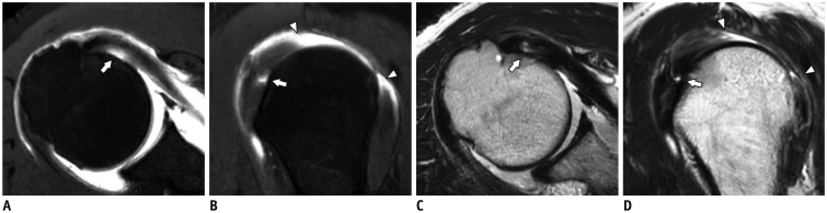
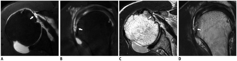
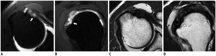
Objective: To compare the T1-weighted spectral presaturation with inversion-recovery sequences (T1 SPIR) with T2-weighted turbo spin-echo sequences (T2 TSE) on 3T magnetic resonance arthrography (MRA) in the evaluation of the subscapularis (SSC) tendon tear with arthroscopic findings as the reference standard.
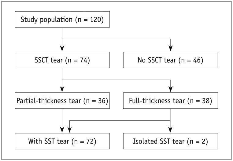
Materials and methods: This retrospective study included 120 consecutive patients who had undergone MRA within 3 months between April and December 2015. Two musculoskeletal radiologists blinded to the arthroscopic results evaluated T1 SPIR and T2 TSE images in separate sessions for the integrity of the SSC tendon, examining normal/articular-surface partial-thickness tear (PTTa)/full-thickness tear (FTT). Diagnostic performance of T1 SPIR and T2 TSE was calculated with arthroscopic results as the reference standard, and sensitivity, specificity, and accuracy were compared using the McNemar test. Interobserver agreement was measured with kappa (κ) statistics.
Results: There were 74 SSC tendon tears (36 PTTa and 38 FTT) confirmed by arthroscopy. Significant differences were found in the sensitivity and accuracy between T1 SPIR and T2 TSE using the McNemar test, with respective rates of 95.9-94.6% vs. 71.6-75.7% and 90.8-91.7% vs. 79.2-83.3% for detecting tear; 55.3% vs. 31.6-34.2% and 85.8% vs. 78.3-79.2%, respectively, for FTT; and 91.7-97.2% vs. 58.3-61.1% and 89% vs. 78-79.3%, respectively, for PTTa. Interobserver agreement for T1 SPIR was almost perfect for T1 SPIR (κ = 0.839) and substantial for T2 TSE (κ = 0.769).
Conclusion: T1-weighted spectral presaturation with inversion-recovery sequences is more sensitive and accurate compared to T2 TSE in detecting SSC tendon tear on 3T MRA.
Keywords: Arthroscopy; Magnetic resonance arthrography; Magnetic resonance imaging; Subscapularis; Tear; Tendon.
Figures
References
-
- Blasier RB, Soslowsky LJ, Malicky DM, Palmer ML. Posterior glenohumeral subluxation: active and passive stabilization in a biomechanical model. J Bone Joint Surg Am. 1997;79:433–440. - PubMed
-
- Halder A, Zobitz ME, Schultz E, An KN. Structural properties of the subscapularis tendon. J Orthop Res. 2000;18:829–834. - PubMed
-
- Abboud JA, Soslowsky LJ. Interplay of the static and dynamic restraints in glenohumeral instability. Clin Orthop Relat Res. 2002:48–57. - PubMed
-
- Morag Y, Jamadar DA, Miller B, Dong Q, Jacobson JA. The subscapularis: anatomy, injury, and imaging. Skeletal Radiol. 2011;40:255–269. - PubMed
-
- Bergin D, Parker L, Zoga A, Morrison W. Abnormalities on MRI of the subscapularis tendon in the presence of a full-thickness supraspinatus tendon tear. AJR Am J Roentgenol. 2006;186:454–459. - PubMed
MeSH terms
LinkOut - more resources
Full Text Sources
Other Literature Sources
Miscellaneous
