. 2018 Feb 22:11:51.
doi: 10.3389/fnmol.2018.00051. eCollection 2018.

Acute Down-regulation of BDNF Signaling Does Not Replicate Exacerbated Amyloid-β Levels and Cognitive Impairment Induced by Cholinergic Basal Forebrain Lesion

Affiliations

Acute Down-regulation of BDNF Signaling Does Not Replicate Exacerbated Amyloid-β Levels and Cognitive Impairment Induced by Cholinergic Basal Forebrain Lesion

Marion T Turnbull et al. Front Mol Neurosci. .

Abstract

Degeneration of basal forebrain cholinergic neurons (BFCNs) precedes hippocampal degeneration and pathological amyloid-beta (Aβ) accumulation, and underpins the development of cognitive dysfunction in sporadic Alzheimer's disease (AD). We hypothesized that degeneration of BFCNs causes a decrease in neurotrophin levels in innervated brain areas, which in turn promotes the development of Aβ pathology and cognitive impairment. Here we show that lesion of septo-hippocampal BFCNs in a pre-symptomatic transgenic amyloid AD mouse model (APP/PS1 mice) increases soluble Aβ levels in the hippocampus, and induces cognitive deficits in a spatial memory task that are not seen in either unlesioned APP/PS1 or non-transgenic littermate control mice. Furthermore, the BFCN lesion results in decreased levels of brain-derived neurotrophic factor (BDNF). However, viral knockdown of neuronal BDNF in the hippocampus of APP/PS1 mice (in the absence of BFCN loss) neither increased the level of Aβ nor caused cognitive deficits. These results suggest that the cognitive decline and Aβ pathology induced by BFCN loss occur independent of dysfunctional neuronal BDNF signaling, and may therefore be directly underpinned by reduced cholinergic neurotransmission.

Keywords: APP/PS1 transgenic mouse; Alzheimer’s disease; amyloid-β; basal forebrain; brain-derived neurotrophic factor; cholinergic neuron.


Figures

Figure 1
Figure 1
p75-saporin toxin injection into the basal forebrain selectively lesions cholinergic neurons. (A) Representative schematic of the paradigm indicating the age and time of surgery and experimental manipulations. (B) Diagram of a murine brain in the sagittal plane (left), with a red box indicating the location of the basal forebrain, and the coronal plane (right), indicating the location of the saporin injection site in the medial septum region of the basal forebrain (in red). (C) Representative photomicrographs of coronal brain sections at the level of the basal forebrain, immunostained for a cholinergic neuron marker (anti-ChAT) 1 month after microinjection of IgG-saporin (control) or p75-saporin (lesion) into the medial septum. (D) Quantification of ChAT- and (E) parvalbumin (PARV)-positive neurons in the basal forebrain of basal forebrain cholinergic neuron (BFCN)-lesioned and control transgenic mice. The number of animals per condition are indicated within the bars of the graphs. *p < 0.05, unpaired t-test. (F) Representative western blots of ChAT protein levels in the hippocampus of naïve, unlesioned (control) and BFCN-lesioned amyloid precursor protein (APP)/PS1 transgenic mice. Data are presented as mean ± SEM.
Figure 2
Figure 2
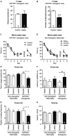
Memory retention is impaired in a Morris water maze task following BFCN lesion in APP/PS1 mice. Graphs of total distance traveled (A) and number of maze arm alternations (B) of basal forebrain-lesioned (p75-saporin) APP/PS1 mice and their non-lesioned (IgG-saporin) transgenic controls (mean ± SEM, ***p < 0.001, unpaired t-test). Graph of the escape latency (time to find the hidden platform) averaged per day in an 8 day Morris water maze task of spatial learning of (C) basal forebrain-lesioned (p75-saporin) APP/PS1 mice and their non-lesioned (IgG-saporin) transgenic controls, and (D) basal forebrain-lesioned (p75-saporin) non-lesioned (IgG-saporin) and naïve non-transgenic littermates of the same age. A probe trial in which the platform was removed was performed on day 6. The platform location was then changed for testing on days 7 and 8. Two-way analysis of variance (ANOVA) with Tukey’s post hoc test, *p < 0.05. (E) Quantification of time spent in the target quadrant, (F) latency to the platform, and (G) platform crossing frequency during the probe trial on day 6, between basal forebrain-lesioned and non-lesioned APP/PS1 transgenic mice and basal forebrain-lesioned, non-lesioned, and naïve non-transgenic littermate controls. (H) Quantification of velocity during the acquisition stage. Data are presented as mean ± SEM with sample size indicated in the bars. One-way ANOVA with Tukey’s post hoc, *p < 0.05, ns, not significant.
Figure 3
Figure 3
Total Aβ42 levels increase in the hippocampus following BFCN lesion. (A) Quantification of total Aβ42 levels by ELISA from hippocampal lysates of control and basal forebrain-lesioned APP/PS1 mice. Quantification of (B) Aβ plaque number with Thioflavin S staining per μm2 and (C) plaque size across the complete hippocampus of basal forebrain-lesioned and control mice. Loss of BFCNs had no effect on plaque number or size in the hippocampus but resulted in an increase in total Aβ42 levels in the hippocampus. Data are presented as mean ± SEM, with sample size indicated in the bars. Unpaired t-test, *p < 0.05. (D) Representative fluorescence images of amyloid plaques (visualized with Thioflavin S staining) in the hippocampus of APP/PS1 transgenic mice with IgG-saporin injection (control) or p75-saporin injection (lesion) into the basal forebrain. Plaques are indicated by white arrows and a magnified view of a plaque is visible in the top right corner.
Figure 4
Figure 4
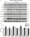
Tau phosphorylation does not change in the hippocampus following BFCN lesion. (A) Representative western blots of the soluble tau fraction of hippocampal homogenates from naïve tau knockout mice, and naïve, control (IgG-saporin) and basal forebrain-lesioned (p75-saporin) APP/PS1 mice probed for the pan-tau marker Tau5 and the tau hyperphosphorylation epitopes AT8, AT180, AT270, pSer235, pSer262 and pSer422. Each tau band is compared to its respective GAPDH loading control band. (B) Quantification of tau protein bands from the soluble tau fraction. No significant differences (two-way ANOVA) in hyperphosphorylated tau epitopes were observed between naïve, control and basal forebrain-lesioned APP/PS1 mice. Bands were normalized to the GAPDH loading control levels and the levels are plotted relative to those of control APP/PS1 mice. Data are presented as the mean ± SEM, with the sample size indicated in the bars.
Figure 5
Figure 5
Brain-derived neurotrophic factor (BDNF) protein and TrkB receptor signaling are reduced following BFCN lesion. (A) Quantification of BDNF protein levels by ELISA from hippocampal lysates of naïve wild-type mice and control and basal forebrain-lesioned APP/PS1 mice. Loss of BFCNs resulted in a decrease in the BDNF protein level. (B) Representative western blots of hippocampal homogenates from control (IgG-saporin) and basal forebrain-lesioned (p75-saporin) APP/PS1 mice probed for phosphorylated TrkB (pTrkB) and total TrkB. Each band is compared to its respective GAPDH loading control band. (C) Quantification of pTrkB and TrkB bands expressed as a percentage of the levels in control mice. Loss of the cholinergic projection to the hippocampus resulted in a reduction in phosphorylated/active TrkB receptor. Data are presented as mean ± SEM, with the sample size indicated in the bars. One-way ANOVA, **p < 0.01, ***p < 0.001.
Figure 6
Figure 6
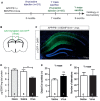
A selective reduction in the hippocampal BDNF level does not alter working memory. (A) Representative schematic of the paradigm indicating the age and time of surgery and the experimental manipulations. (B) Diagram of a murine brain in the coronal plane with green lines indicating the location of the stereotaxic injection of virus (AAV-Synapsin-Cre-GFP). (C) Representative fluorescence image of a hippocampal cross-section demonstrating the spatial distribution of the virus carrying a GFP tag (in green) after stereotaxic injection into an APP/PS1 × BDNFfl/wt mouse. (D) Quantification of the BDNF protein levels (pg/mg total protein) by ELISA from anterior hippocampal lysates of naïve (gray bars), saline-injected and virus-injected APP/PS1 × BDNFfl/wt mice. Stereotaxic injection of AAV-Synapsin-Cre-GFP into the hippocampus of mice carrying a floxed BDNF allele resulted in a decrease in BDNF protein. One-way ANOVA, **p < 0.01. Graphs of total distance traveled (E) and the number of maze arm alternations (F) of APP/PS1 × BDNFfl/wt mice injected with a saline control or AAV Synapsin-Cre virus (unpaired t-test), ns, not significant.
Figure 7
Figure 7
A selective reduction in the hippocampal BDNF level does not alter the level of Aβ or affect amyloid pathology. (A) Representative fluorescence images of the hippocampus of APP/PS1 × BDNFfl/wt mice injected with a saline control or virus. Amyloid plaques were labeled with an Aβ antibody (red, arrowed), and cells infected with the virus were labeled with a GFP antibody (green). Quantification of the number of amyloid plaques in (B) the CA1/CA2 and (C) the dentate gyrus subregions of the hippocampus of APP/PS1 × BDNFfl/wt mice injected with a saline control or virus. (D) Quantification of total Aβ42 levels by ELISA from anterior hippocampal lysates of saline- and virus-injected APP/PS1 × BDNFfl/wt mice. A reduction in the BDNF protein level in the hippocampus did not affect the number of amyloid plaques, or the level of soluble Aβ. Data are presented as mean ± SEM, with the sample size indicated in the bars.

Similar articles

Cited by

References

    1. Al-Onaizi M. A., Parfitt G. M., Kolisnyk B., Law C. S., Guzman M. S., Barros D. M., et al. . (2017). Regulation of cognitive processing by hippocampal cholinergic tone. Cereb. Cortex 27, 1615–1628. 10.1093/cercor/bhv349 - DOI - PubMed
    1. Arancibia S., Silhol M., Moulière F., Meffre J., Höllinger I., Maurice T., et al. . (2008). Protective effect of BDNF against β-amyloid induced neurotoxicity in vitro and in vivo in rats. Neurobiol. Dis. 31, 316–326. 10.1016/j.nbd.2008.05.012 - DOI - PubMed
    1. Blurton-Jones M., Kitazawa M., Martinez-Coria H., Castello N. A., Müller F. J., Loring J. F., et al. . (2009). Neural stem cells improve cognition via BDNF in a transgenic model of Alzheimer disease. Proc. Natl. Acad. Sci. U S A 106, 13594–13599. 10.1073/pnas.0901402106 - DOI - PMC - PubMed
    1. Boskovic Z., Alfonsi F., Rumballe B. A., Fonseka S., Windels F., Coulson E. J. (2014). The role of p75NTR in cholinergic basal forebrain structure and function. J. Neurosci. 34, 13033–13038. 10.1523/JNEUROSCI.2364-14.2014 - DOI - PMC - PubMed
    1. Burgess B. L., Mcisaac S. A., Naus K. E., Chan J. Y., Tansley G. H., Yang J., et al. . (2006). Elevated plasma triglyceride levels precede amyloid deposition in Alzheimer’s disease mouse models with abundant Aβ in plasma. Neurobiol. Dis. 24, 114–127. 10.1016/j.nbd.2006.06.007 - DOI - PubMed

LinkOut - more resources