. 2018 Feb 19:9:272.
doi: 10.3389/fimmu.2018.00272. eCollection 2018.

Extracellular Vesicles Released from Mycobacterium tuberculosis-Infected Neutrophils Promote Macrophage Autophagy and Decrease Intracellular Mycobacterial Survival

Affiliations

Extracellular Vesicles Released from Mycobacterium tuberculosis-Infected Neutrophils Promote Macrophage Autophagy and Decrease Intracellular Mycobacterial Survival

Violeta D Alvarez-Jiménez et al. Front Immunol. .

Abstract

Tuberculosis is an infectious disease caused by Mycobacterium tuberculosis (Mtb). In the lungs, macrophages and neutrophils are the first immune cells that have contact with the infecting mycobacteria. Neutrophils are phagocytic cells that kill microorganisms through several mechanisms, which include the lytic enzymes and antimicrobial peptides that are found in their lysosomes, and the production of reactive oxygen species. Neutrophils also release extracellular vesicles (EVs) (100-1,000 nm in diameter) to the extracellular milieu; these EVs consist of a lipid bilayer surrounding a hydrophilic core and participate in intercellular communication. We previously demonstrated that human neutrophils infected in vitro with Mtb H37Rv release EVs (EV-TB), but the effect of these EVs on other cells relevant for the control of Mtb infection, such as macrophages, has not been completely analyzed. In this study, we characterized the EVs produced by non-stimulated human neutrophils (EV-NS), and the EVs produced by neutrophils stimulated with an activator (PMA), a peptide derived from bacterial proteins (fMLF) or Mtb, and observed that the four EVs differed in their size. Ligands for toll-like receptor (TLR) 2/6 were detected in EV-TB, and these EVs favored a modest increase in the expression of the co-stimulatory molecules CD80, a higher expression of CD86, and the production of higher amounts of TNF-α and IL-6, and of lower amounts of TGF-β, in autologous human macrophages, compared with the other EVs. EV-TB reduced the amount of intracellular Mtb in macrophages, and increased superoxide anion production in these cells. TLR2/6 ligation and superoxide anion production are known inducers of autophagy; accordingly, we found that EV-TB induced higher expression of the autophagy-related marker LC3-II in macrophages, and the co-localization of LC3-II with Mtb inside infected macrophages. The intracellular mycobacterial load increased when autophagy was inhibited with wortmannin in these cells. In conclusion, our results demonstrate that neutrophils produce different EVs in response to diverse activators, and that EV-TB activate macrophages and promote the clearance of intracellular Mtb through early superoxide anion production and autophagy induction, which is a novel role for neutrophil-derived EVs in the immune response to Mtb.

Keywords: autophagy; extracellular vesicles; macrophage; neutrophils; tuberculosis.


Figures
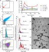
Figure 1
Figure 1
Neutrophil extracellular vesicles (EVs) that are induced by Mycobacterium tuberculosis (Mtb) have different characteristics than neutrophil EVs induced by PMA or fMLF. EVs were derived from neutrophils that were left with medium alone (EV-NS) or stimulated with PMA (EV-PMA), fMLF (EV-fMLF), or with Mtb (EV-TB) for the indicated times. (A) Gating strategy for the flow cytometry analysis of EVs. (a) Side scatter (SSC-A) of MegaMix® beads of different sizes, which allowed the differentiation of EVs from the electronic noise. (b) SSC-A of neutrophil-derived EVs, (c) EVs derived from neutrophils stained with CellVue Jade, and (d) analysis of the expression of CD35 and annexin V on neutrophil-derived EVs. (B) Percentage of CD35+, annexin V+ EVs derived from neutrophils activated with different stimuli. Data points represent mean and SEM from four independent experiments and were analyzed (at each time point) with Kruskal–Wallis test with Dunn’s posttest. Asterisks on the graph represent significant differences between EV-NS and EV-PMA, EV-fMLF, or EV-TB (*P < 0.05 and **P < 0.01). (C) Nanoparticle tracking analysis (NTA) of EVs derived from neutrophils activated with different stimuli. Three measurements were made from each sample. A result representative of four independent experiments is shown. (D) Transmission electron microscopy (TEM) of EVs derived from neutrophil activated with different stimuli. The red bars indicate 100 nm, and the white arrows indicate the largest EVs. The images are representative of three independent experiments.
Figure 2
Figure 2
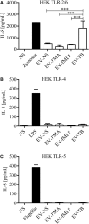
Extracellular vesicles (EVs) derived from Mycobacterium tuberculosis (Mtb)-infected neutrophils (EV-TB) contain ligands for toll-like receptors (TLR) 2/6. HEK cells expressing TLR2/6 (A), TLR4 (B), or TLR5 (C) were stimulated with EVs that were produced by non-stimulated neutrophils (EV-NS) or by neutrophils stimulated with PMA (EV-PMA), fMLF (EV-fMLF), or Mtb (EV-TB) for 30 min. Zymosan (TLR 2/6), lipopolysaccharide (LPS) (TLR4) and flagellin (TLR5) were used as positive controls, as indicated, and non-stimulated cells were used as negative controls (NS). After 24 h, supernatants were collected, and IL-8 was quantified. Data points represent mean and SD from three independent experiments and were analyzed with one-way ANOVA followed by Tukey’s test (***P < 0.001).
Figure 3
Figure 3
Neutrophil-derived extracellular vesicles (EVs) that are induced by Mycobacterium tuberculosis (Mtb) induce the production of TNF-α and IL-6 and IL-10 by macrophages. Macrophages were stimulated for 24 h with EVs that were produced by non-stimulated neutrophils (EV-NS), or by neutrophils stimulated with PMA (EV-PMA), fMLF (EV-fMLF), or Mtb (EV-TB) for 30 min. As controls, the macrophages were left with medium alone (NS) or were infected with Mtb. TNF-α (A), IL-6 (B), IL-1β (C), IL-10 (D), and TGF-β (E) were measured in the supernatants. The graphs represent the results obtained with cells from five different healthy volunteers and were analyzed with one-way ANOVA followed by Tukey’s test (*P < 0.05, **P < 0.01, and ***P < 0.001).
Figure 4
Figure 4
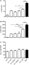
Effect of neutrophil-derived extracellular vesicles (EVs) on the expression of costimulatory molecules in macrophages. Macrophages were stimulated for 24 h with EVs that were produced by non-stimulated neutrophils (EV-NS), or by neutrophils stimulated with PMA (EV-PMA), fMLF (EV-fMLF), or Mycobacterium tuberculosis (Mtb) (EV-TB) for 30 min. As controls, the macrophages were left with medium alone (NS) or were infected with Mtb. The expression of CD80 (A), CD86 (B), and HLA-DR (C) was analyzed by flow cytometry. The graphs represent the results obtained with cells from five different healthy volunteers, and were analyzed with Kruskal–Wallis test and Dunn’s posttest (*P < 0.05, **P < 0.01, and ***P < 0.001).
Figure 5
Figure 5
EV-TB reduce the amount of intracellular Mycobacterium tuberculosis (Mtb) in macrophages. Macrophages were infected with Mtb at a multiplicity of infection (MOI) of 10 for 2 h. Extracellular bacteria were eliminated with amikacin, and the cells were left untreated (IM) or stimulated with the four types of extracellular vesicles (EVs). The EVs were produced by non-stimulated neutrophils (EV-NS), or by neutrophils stimulated with PMA (EV-PMA), fMLF (EV-fMLF), or Mtb at an MOI of 10 (EV-TB) for 30 min. After 4 h of stimulation with EVs, macrophages were washed with PBS and incubated for a total of 24 and 48 h after infection. Finally, IM were lysed, and intracellular bacteria were evaluated through CFU by performing serial dilutions of macrophage lysates. Data points represent the mean and SD from three independent experiments and were analyzed (at each time point) with one-way ANOVA followed by Tukey’s test. Asterisks on the graph represent significant differences between IM and IM plus each EV (*P < 0.05, **P < 0.01, and ***P < 0.001). Abbreviation: IM, infected macrophages.
Figure 6
Figure 6
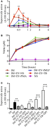
EV-TB induce the production of superoxide anion in Mycobacterium tuberculosis (Mtb)-IM. Macrophages were infected with Mtb for 2 h. Extracellular bacteria were eliminated, and macrophages were left untreated (IM), or incubated with extracellular vesicles (EVs) (EV-NS, EV-PMA, EV-fMLF, or EV-TB) for 4 h. Un-IM were used as negative controls (NS). (A) Superoxide anion was measured with the nitro blue tetrazolium method. The graph represents the change in optical density at 620 nm ± SD of stimulated cells, compared with untreated cells. (B) NO was measured with the Griess method. Data from three independent experiments were analyzed (at each time point) with one-way ANOVA followed by Tukey’s test. Asterisks on the graph represent significant differences between IM and IM plus each EV and the bar in the superoxide anion graph represent significant differences between EV-TB and each EV. (C) NADPH oxidase inhibition with DPI. The graph represents the change in optical density at 620 nm ± SD of stimulated cells, compared with untreated cells. Results were obtained with cells from three different healthy volunteers and were analyzed with one-way ANOVA followed by Tukey’s test (*P < 0.05, **P < 0.01, and ***P < 0.001). Abbreviation: IM, infected macrophages.
Figure 7
Figure 7
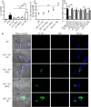
EV-TB-induced autophagy contributes to Mycobacterium tuberculosis (Mtb) elimination in Mtb-IM. (A) Macrophages were incubated with extracellular vesicles (EVs) (EV-NS, EV-PMA, EV-fMLF, or EV-TB) for 4 h. Non-stimulated cells were used as negative control (NS). As a positive control for autophagy induction, macrophages were treated with peptidoglycan (PGN) for 4 h. The cells were then stained with anti-LC3-II (green) and DAPI (blue) and examined in a confocal microscope to determine LC3-II mean fluorescence intensity (MFI). Data points represent the mean and SEM from three independent experiments and were analyzed with Kruskal–Wallis test and Dunn’s posttest (*P < 0.05 and **P < 0.01). (B) Macrophages were infected with Mtb previously stained with CellVue Maroon (red) for 15 min. After this incubation, the cells were left untreated (IM), or were incubated with EVs (EV-NS, EV-PMA, EV-fMLF, or EV-TB) for 4 h. The cells were then stained with anti-LC3-II (green) and DAPI (blue) and examined in a confocal microscope. The images are representative of three independent experiments. (C) Percentage of cells with co-localization of LC3-II and Mtb was calculated. The medians with the interquartile ranges are shown (n = 3) and were analyzed with Kruskal–Wallis test and Dunn’s posttest (**P < 0.01). (D) Percentage of intracellular Mtb survival. Macrophages were infected with Mtb for 2 h. Extracellular bacteria were eliminated, and cells were incubated with EVs (EV-NS, EV-PMA, EV-fMLF, or EV-TB) for 4 h, with or without wortmannin (autophagy inhibitor); IM were also treated with rapamycin (autophagy positive control) or wortmannin. The cells were washed with PBS and incubated for 4 h. The cells were then lysed, and intracellular bacteria were evaluated through CFU by performing serial dilutions of macrophage lysates. The percentage of Mtb survival was calculated for each condition, with Mtb-IM considered as 100%. Data points represent the mean and SEM from three independent experiments and were analyzed with Kruskal–Wallis test and Dunn’s posttest (*P < 0.05, **P < 0.01, and ***P < 0.001). Abbreviation: IM, infected macrophages.

References

    1. Schafer G, Jacobs M, Wilkinson RJ, Brown GD. Non-opsonic recognition of Mycobacterium tuberculosis by phagocytes. J Innate Immun (2009) 1(3):231–43. 10.1159/000173703 - DOI - PMC - PubMed
    1. Tsao TC, Hong J, Huang C, Yang P, Liao SK, Chang KS. Increased TNF-alpha, IL-1 beta and IL-6 levels in the bronchoalveolar lavage fluid with the upregulation of their mRNA in macrophages lavaged from patients with active pulmonary tuberculosis. Tuber Lung Dis (1999) 79(5):279–85. 10.1054/tuld.1999.0215 - DOI - PubMed
    1. D’Avila H, Roque NR, Cardoso RM, Castro-Faria-Neto HC, Melo RC, Bozza PT. Neutrophils recruited to the site of Mycobacterium bovis BCG infection undergo apoptosis and modulate lipid body biogenesis and prostaglandin E production by macrophages. Cell Microbiol (2008) 10(12):2589–604. 10.1111/j.1462-5822.2008.01233.x - DOI - PubMed
    1. Eruslanov EB, Lyadova IV, Kondratieva TK, Majorov KB, Scheglov IV, Orlova MO, et al. Neutrophil responses to Mycobacterium tuberculosis infection in genetically susceptible and resistant mice. Infect Immun (2005) 73(3):1744–53. 10.1128/IAI.73.3.1744-1753.2005 - DOI - PMC - PubMed
    1. Eum SY, Kong JH, Hong MS, Lee YJ, Kim JH, Hwang SH, et al. Neutrophils are the predominant infected phagocytic cells in the airways of patients with active pulmonary TB. Chest (2010) 137(1):122–8. 10.1378/chest.09-0903 - DOI - PMC - PubMed

Publication types