Review
. 2018 Feb 19:9:292.
doi: 10.3389/fimmu.2018.00292. eCollection 2018.

Adrenomedullin and Adrenomedullin-Targeted Therapy As Treatment Strategies Relevant for Sepsis

Affiliations
Review

Adrenomedullin and Adrenomedullin-Targeted Therapy As Treatment Strategies Relevant for Sepsis

Christopher Geven et al. Front Immunol. .

Abstract

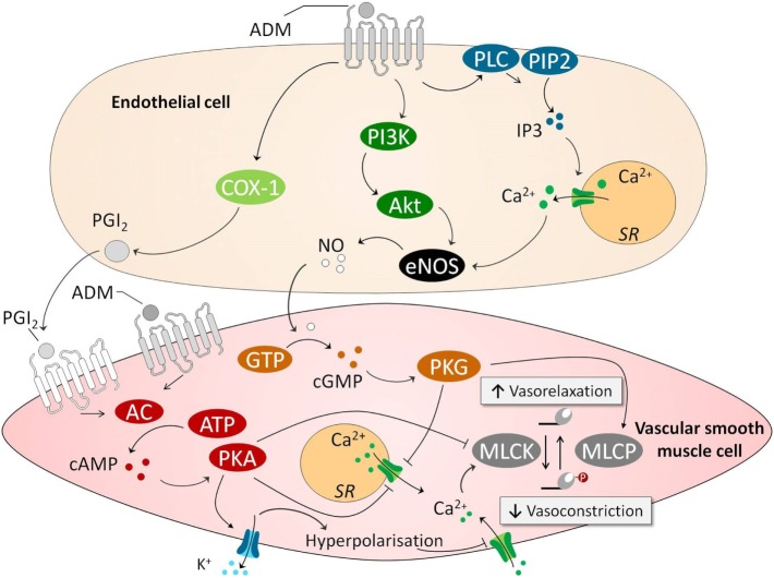
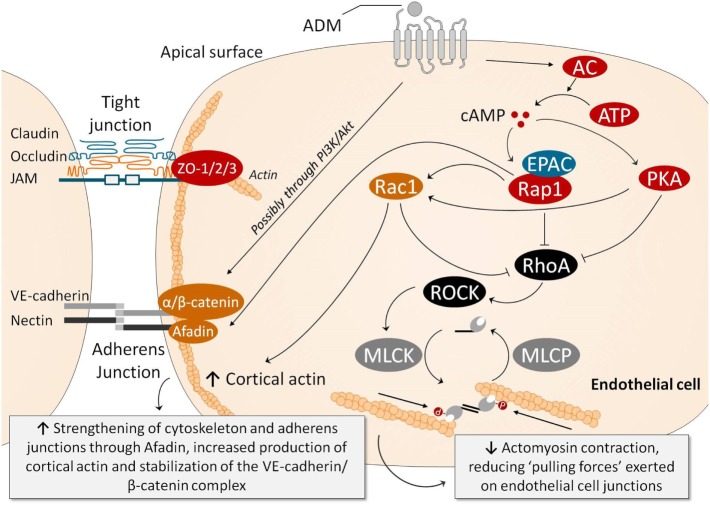
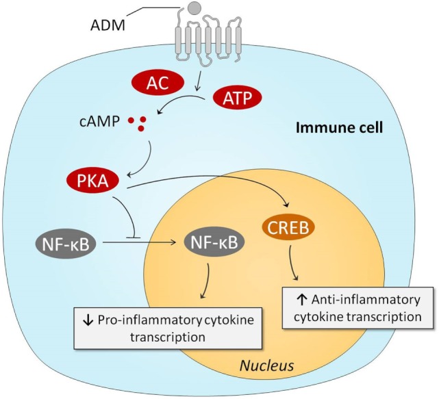
Sepsis remains a major medical challenge, for which, apart from improvements in supportive care, treatment has not relevantly changed over the last few decades. Vasodilation and vascular leakage play a pivotal role in the development of septic shock, with vascular leakage being caused by disrupted endothelial integrity. Adrenomedullin (ADM), a free circulating peptide involved in regulation of endothelial barrier function and vascular tone, is implicated in the pathophysiology of sepsis. ADM levels are increased during sepsis, and correlate with extent of vasodilation, as well as with disease severity and mortality. In vitro and preclinical in vivo data show that administration of ADM exerts anti-inflammatory, antimicrobial, and protective effects on endothelial barrier function during sepsis, but other work suggests that it may also decrease blood pressure, which could be detrimental for patients with septic shock. Work has been carried out to negate ADMs putative negative effects, while preserving or even potentiating its beneficial actions. Preclinical studies have demonstrated that the use of antibodies that bind to the N-terminus of ADM results in an overall increase of circulating ADM levels and improves sepsis outcome. Similar beneficial effects were obtained using coadministration of ADM and ADM-binding protein-1. It is hypothesized that the mechanism behind the beneficial effects of ADM binding involves prolongation of its half-life and a shift of ADM from the interstitium to the circulation. This in turn results in increased ADM activity in the blood compartment, where it exerts beneficial endothelial barrier-stabilizing effects, whereas its detrimental vasodilatory effects in the interstitium are reduced. Up till now, in vivo data on ADM-targeted treatments in humans are lacking; however, the first study in septic patients with an N-terminus antibody (Adrecizumab) is currently being conducted.

Keywords: adrenomedullin; antibodies; sepsis; septic shock; treatment; vascular barrier function.


Figures

Figure 1
Figure 1
ADM causes vasodilation through endothelium-dependent and endothelium-independent pathways. In an endothelium-independent way, binding of ADM with its receptors on VSMCs increases intracellular cAMP. This leads to subsequent activation PKA, which inhibits smooth muscle cell contraction in several ways. First, PKA opens VSMC potassium channels, causing potassium efflux, leading to membrane potential hyperpolarization and closing of voltage gated calcium channels, reducing intracellular calcium content. Other effects of PKA include inhibition of sarcoplasmatic calcium channel and MLCK. The latter of which is essential for actomyosin contraction. Several endothelium-dependent pathways have been identified. This includes a COX/PGI2 pathway that activates the cAMP pathway in VSMCs. Other involved endothelium-dependent pathways are PI3k/Akt and PLC/IP3, which both activate eNOS which leads to subsequently activation of a cGMP/cGMP-dependent kinase pathway in VSMCs. This pathway leads to activation of MLCP which “inactivates” the myosin light chain, and again lowers levels of calcium by inhibiting sarcoplasmatic calcium channels. Abbreviations: AC, adenylyl cyclase; AKT, protein kinase B; ATP, adenosine triphosphate; Ca2+, calcium; cAMP, cyclic adenosine monophosphate; cGMP, cyclic guanosine monophosphate; COX-1, cyclooxygenase-1; eNOS; endothelial nitric oxide synthase; GTP, guanosine triphosphate; IP3, inositol triphosphate; MLCK, myosin light chain kinase; MLCP, myosin light chain phosphatase; NO, nitric oxide; PGI2, prostacyclin; PI3K, phosphoinositide 3-kinase; PIP2, phosphatidylinositol-4,5-bisphosphate; PKA, protein kinase A; PLC, phospholipase C; SR, sarcoplasmatic reticulum; VSMC, vascular smooth muscle cell; ADM, adrenomedullin.
Figure 2
Figure 2
Several pathways have been identified through which ADM exerts endothelial barrier-stabilizing effects. Ligation of ADM with its receptors elicits a strong increase in intracellular cAMP in endothelial cells (ECs), which subsequently activates PKA and, through activation of EPAC, Rap1. PKA and Rap1 inhibit RhoA/ROCK, which results in reduced myosin light chain phosphorylation, decreasing actomyosin contraction (i.e., the “pulling forces” exerted on the EC junctions). Rap1 also promotes junctional adhesiveness via Afadin, strengthening junctional tightening by mediating attachment of AJs and the actin cytoskeleton. PKA also increases cortical actin formation through Rac1, which promotes cell–cell stability and cell–matrix adhesion by its connection to tight and AJs. Moreover, Rac1 is also able to inhibit RhoA, decreasing myosin light chain phosphorylation and actomyosin contraction, similar to PKA and Rap1. Ligation of ADM with its receptor is also thought to prevent phosphorylation of VE-cadherin and β-catenin complexes (which would be detrimental for barrier function because phosphorylation of VE-cadherin prevents binding of β-catenin to the cytoplasmic tail of VE-cadherin, and because phosphorylation of β-catenin targets β-catenin for ubiquination and proteasomal degradation), through the PI3K/Akt pathway. Abbreviations: AC, adenylyl cyclase; ADM, adrenomedullin; AJ, adherens junction; ATP, adenosine triphosphate; cAMP, cyclic adenosine monophosphate; cGMP, cyclic guanosine monophosphate; EPAC, exchange factor directly activated by cAMP; MLCK, myosin light chain kinase; MLCP, myosin light chain phosphatase; PI3K/Akt, phosphatidylinositol-4,5-bisphosphate 3-kinase-protein kinase B; PKA, protein kinase A; Rac, Ras-related C3 botulinum toxin substrate 1; Rap1, Ras-related protein-1; ROCK, rho-associated protein kinase; TJ, tight junction; VE-cadherin, vascular endothelial-cadherin; ZO, zonula occludens.
Figure 3
Figure 3
Intracellular mechanisms behind ADM-induced anti-inflammatory effects. Stimulation of the ADM receptors results in increased intracellular cAMP concentrations, which subsequently activate PKA. PKA prevents NF-κB from entering the nucleus, resulting in reduced transcription of pro-inflammatory genes. PKA-induced activation of CREB results in augmented anti-inflammatory transcription of anti-inflammatory cytokines. Abbreviations: AC, adenylyl cyclase; ADM, adrenomedullin; ATP, adenosine triphosphate; cAMP, cyclic adenosine monophosphate; CREB, cAMP response element-binding protein; NF-κB, nuclear factor kappa-light-chain-enhancer of activated B cells; PKA, protein kinase A.

References

    1. Fleischmann C, Scherag A, Adhikari NK, Hartog CS, Tsaganos T, Schlattmann P, et al. Assessment of global incidence and mortality of hospital-treated sepsis. Current estimates and limitations. Am J Respir Crit Care Med (2016) 193(3):259–72.10.1164/rccm.201504-0781OC - DOI - PubMed
    1. Vincent JL, Marshall JC, Namendys-Silva SA, Francois B, Martin-Loeches I, Lipman J, et al. Assessment of the worldwide burden of critical illness: the intensive care over nations (ICON) audit. Lancet Respir Med (2014) 2(5):380–6.10.1016/s2213-2600(14)70061-x - DOI - PubMed
    1. Singer M, Deutschman CS, Seymour CW, Shankar-Hari M, Annane D, Bauer M, et al. The Third International Consensus definitions for sepsis and septic shock (sepsis-3). JAMA (2016) 315(8):801–10.10.1001/jama.2016.0287 - DOI - PMC - PubMed
    1. Abraham E, Singer M. Mechanisms of sepsis-induced organ dysfunction. Crit Care Med (2007) 35(10):2408–16.10.1097/01.CCM.0000282072.56245.91 - DOI - PubMed
    1. Angus DC, van der Poll T. Severe sepsis and septic shock. N Engl J Med (2013) 369(9):840–51.10.1056/NEJMra1208623 - DOI - PubMed