

Differentiating papillary type I RCC from clear cell RCC and oncocytoma: application of whole-lesion volumetric ADC measurement
- PMID: 29520425
- PMCID: PMC8058623
- DOI: 10.1007/s00261-017-1453-4
Differentiating papillary type I RCC from clear cell RCC and oncocytoma: application of whole-lesion volumetric ADC measurement
Abstract
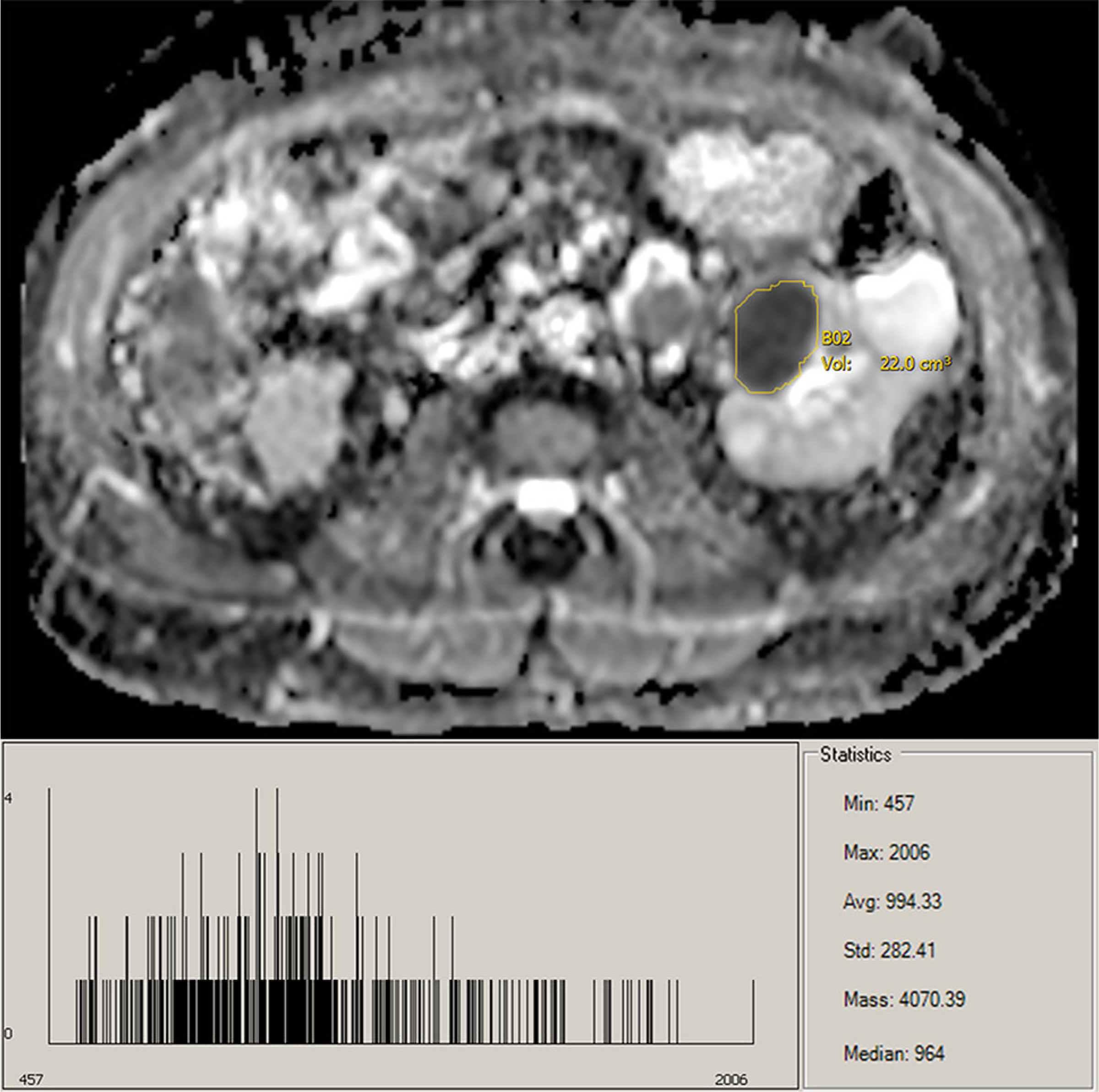
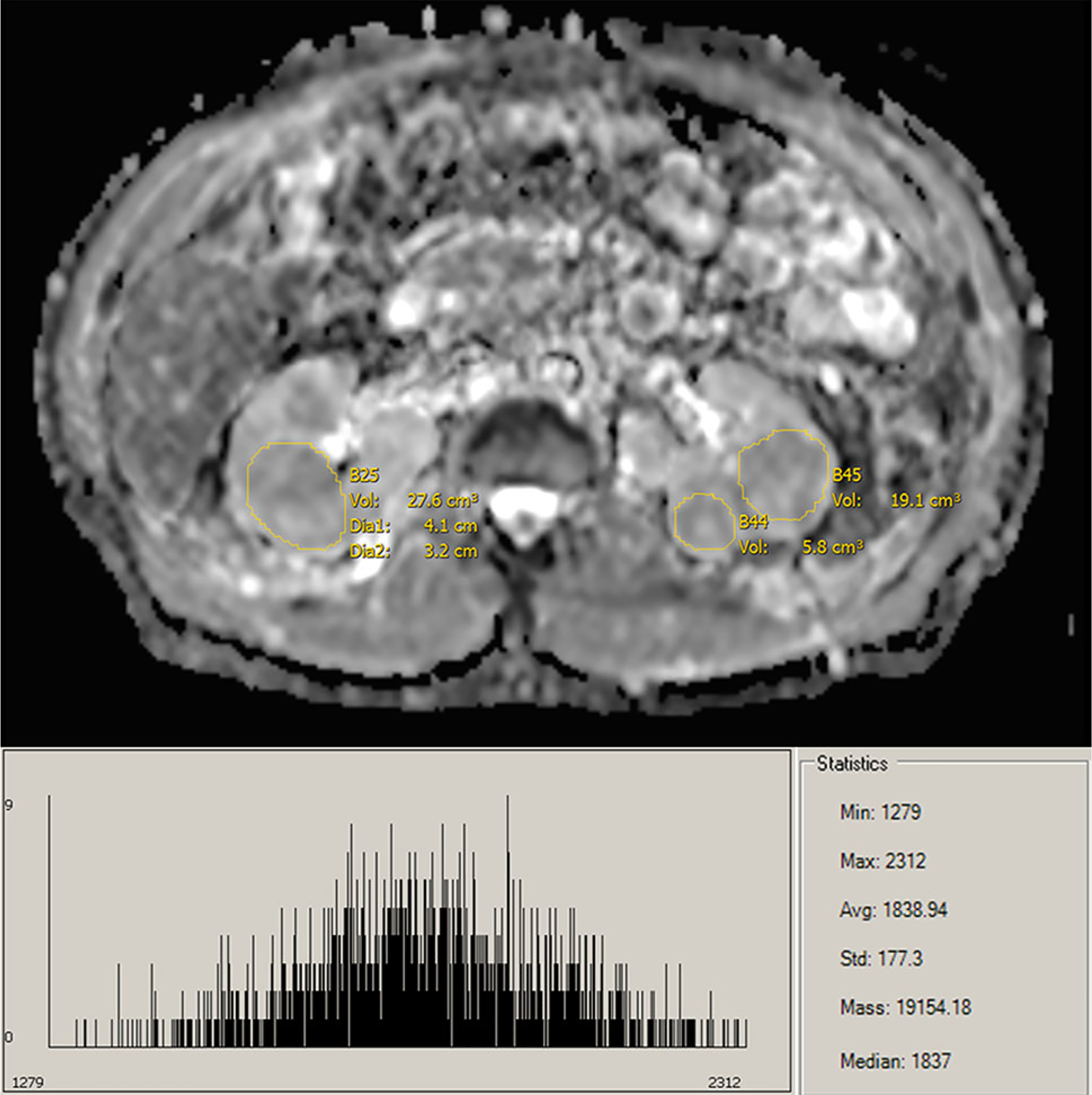
Purpose: To determine whether objective volumetric whole-lesion apparent diffusion coefficient (ADC) distribution analysis improves upon the capabilities of conventional subjective small region-of-interest (ROI) ADC measurements for prediction of renal cell carcinoma (RCC) subtype.
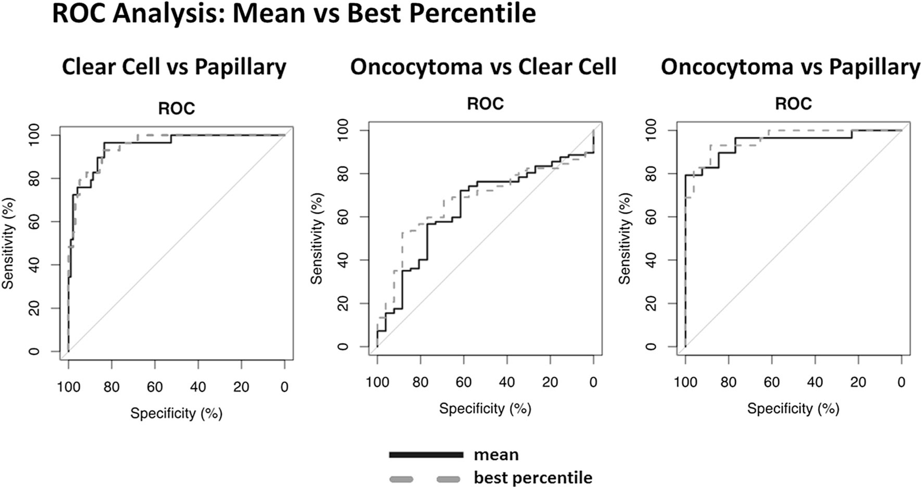
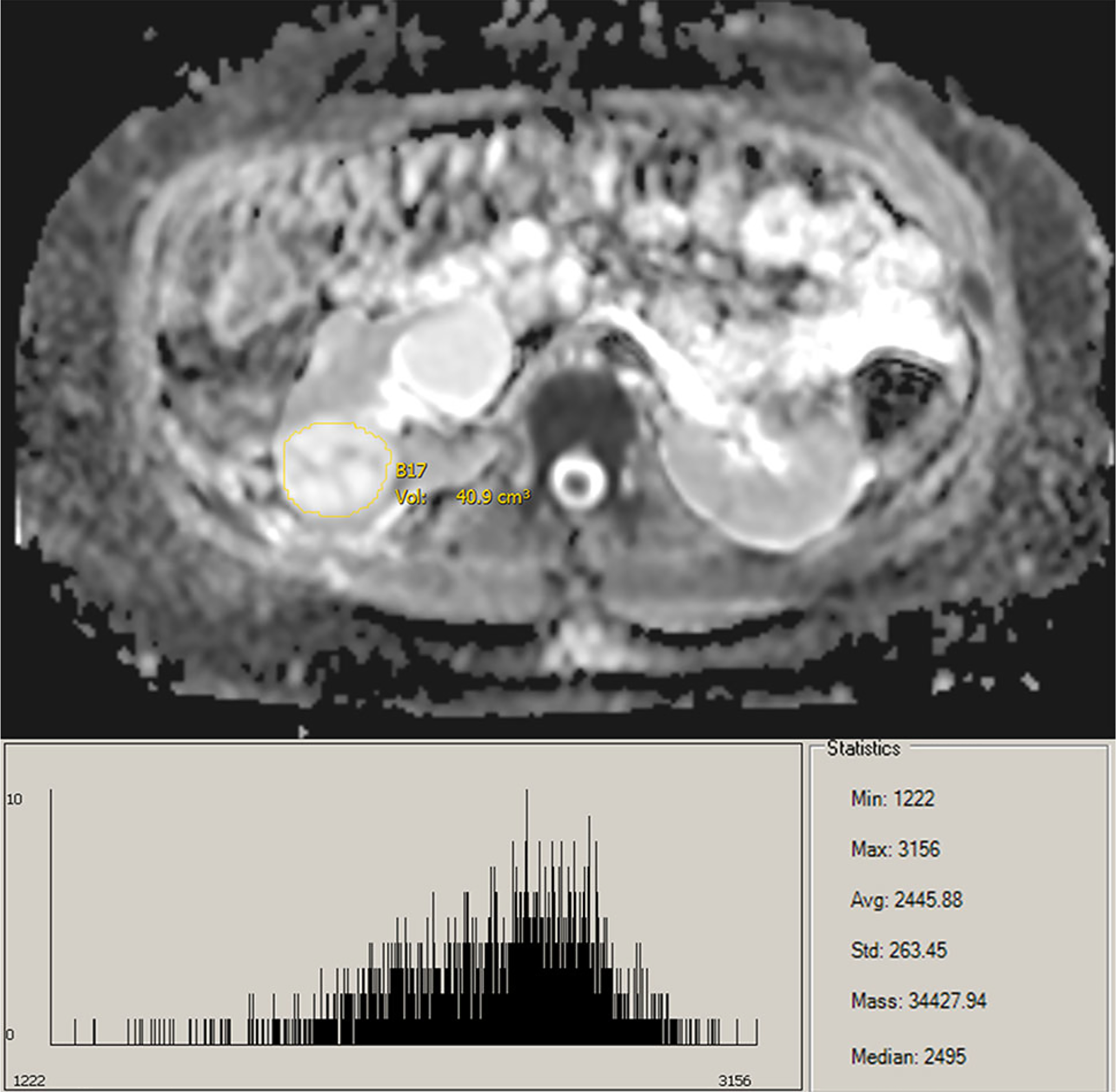
Methods: This IRB-approved study retrospectively enrolled 55 patients (152 tumors). Diffusion-weighted imaging DWI was acquired at b values of 0, 250, and 800 s/mm2 on a 1.5T system (Aera, Siemens Healthcare). Whole-lesion measurements were performed by a research fellow and reviewed by a fellowship-trained radiologist. Mean, median, skewness, kurtosis, and every 5th percentile ADCs were determined from the whole-lesion histogram. Linear mixed models that accounted for within-subject correlation of lesions were used to compare ADCs among RCC subtypes. Receiver-operating characteristic (ROC) curve analysis with optimal cutoff points from the Youden index was used to test the ability of ADCs to differentiate clear cell RCC (ccRCC), papillary RCC (pRCC), and oncocytoma subtypes.
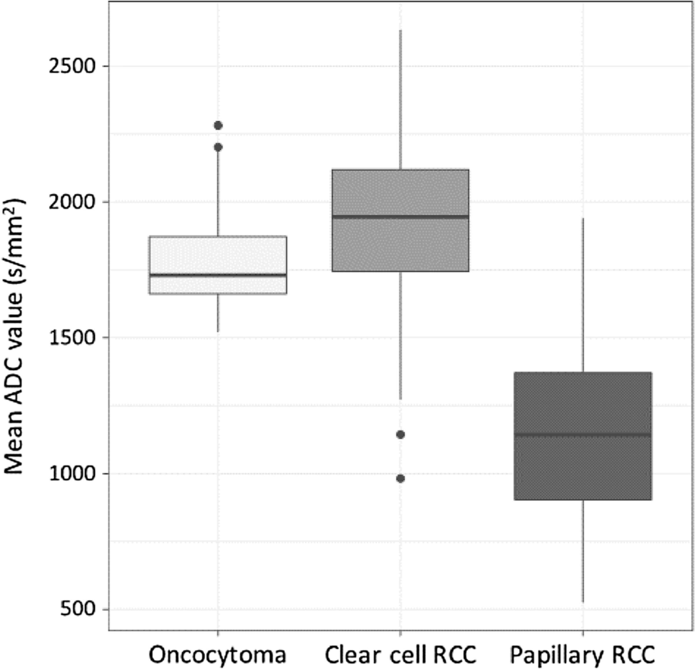
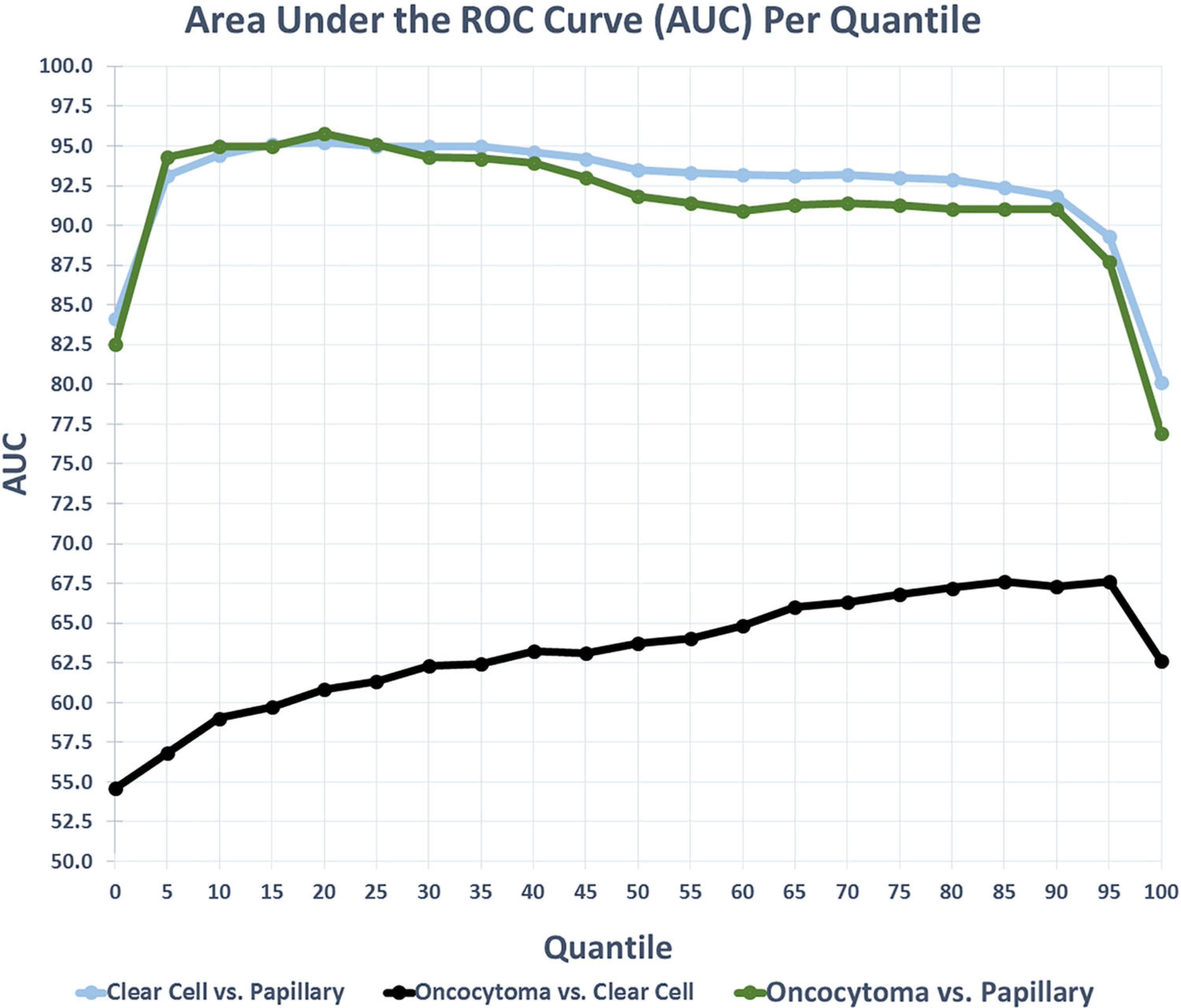
Results: Whole-lesion ADC values were significantly different between pRCC and ccRCC, and between pRCC and oncocytoma, demonstrating strong ability to differentiate subtypes across the quantiles (both P < 0.001). Best percentile ROC analysis demonstrated AUC values of 95.2 for ccRCC vs. pRCC; 67.6 for oncocytoma vs. ccRCC; and 95.8 for oncocytoma vs. pRCC. Best percentile ROC analysis further indicated model sensitivities/specificities of 84.5%/93.1% for ccRCC vs. pRCC; 100.0%/10.3% for oncocytoma vs. ccRCC; and 88.5%/93.1% for oncocytoma vs. pRCC.
Conclusion: The objective methodology of whole-lesion volumetric ADC measurements maintains the sensitivity/specificity of conventional expert-based ROI analysis, provides information on lesion heterogeneity, and reduces observer bias.
Keywords: Apparent diffusion coefficients; Diffusion-weighted imaging; Renal cell carcinoma; Subtype differentiation.
Conflict of interest statement
Compliance with ethical standards
Figures

References
-
- Mytsyk Y, Dutka I, Borys Y, et al. (2017) Renal cell carcinoma: applicability of the apparent coefficient of the diffusion-weighted estimated by MRI for improving their differential diagnosis, histologic subtyping, and differentiation grade. Int urol nephrol 49:215–224 - PubMed
-
- Agnello F, Roy C, Bazille G, et al. (2013) Small solid renal masses: characterization by diffusion-weighted MRI at 3 T. Clin Radiol 68:e301–e308 - PubMed
-
- Zhang H, Gan Q, Wu Y, et al. (2016) Diagnostic performance of diffusion-weighted magnetic resonance imaging in differentiating human renal lesions (benignity or malignancy): a meta-analysis. Abdom Radiol 41:1997–2010 - PubMed
-
- Wang H, Cheng L, Zhang X, et al. (2010) Renal cell carcinoma: diffusion-weighted MR imaging for subtype differentiation at 3.0 T. Radiology 257:135–143 - PubMed
Publication types
MeSH terms
Substances
Grants and funding
LinkOut - more resources
Full Text Sources
Other Literature Sources
Medical
