. 2018 Nov;80(5):1765-1775.
doi: 10.1002/mrm.27166. Epub 2018 Mar 9.

A convolutional neural network to filter artifacts in spectroscopic MRI

Affiliations

A convolutional neural network to filter artifacts in spectroscopic MRI

Saumya S Gurbani et al. Magn Reson Med. 2018 Nov.

Abstract

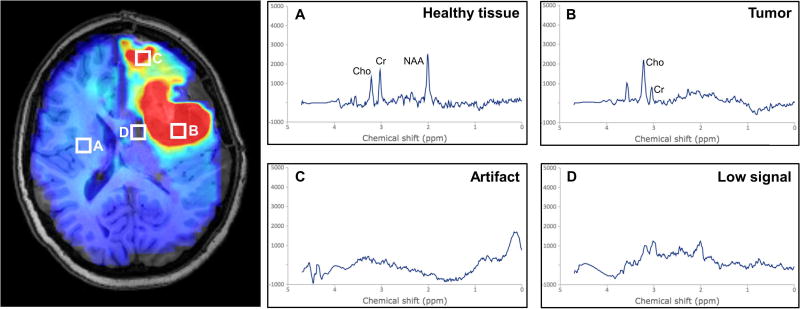
Purpose: Proton MRSI is a noninvasive modality capable of generating volumetric maps of in vivo tissue metabolism without the need for ionizing radiation or injected contrast agent. Magnetic resonance spectroscopic imaging has been shown to be a viable imaging modality for studying several neuropathologies. However, a key hurdle in the routine clinical adoption of MRSI is the presence of spectral artifacts that can arise from a number of sources, possibly leading to false information.

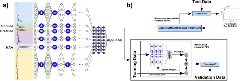
Methods: A deep learning model was developed that was capable of identifying and filtering out poor quality spectra. The core of the model used a tiled convolutional neural network that analyzed frequency-domain spectra to detect artifacts.

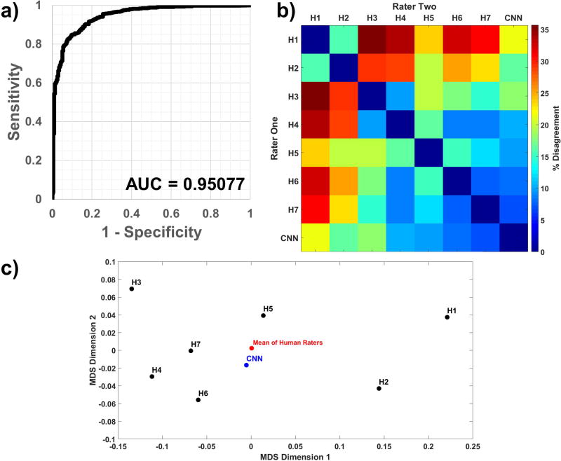
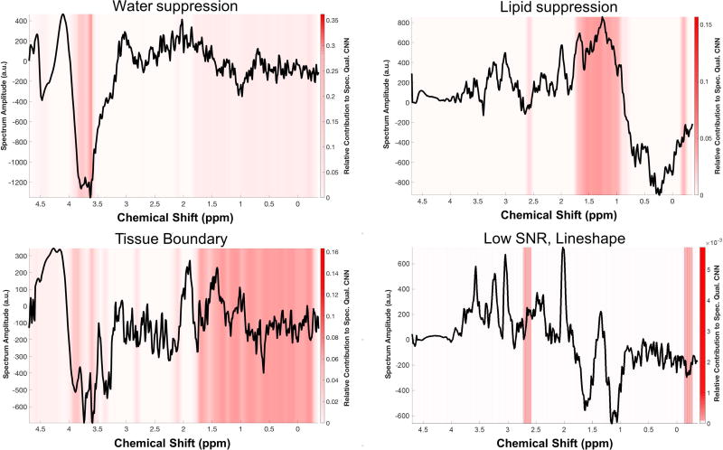
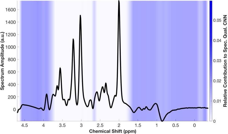
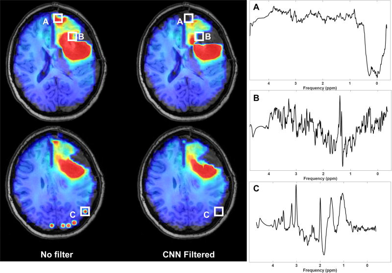
Results: When compared with a panel of MRS experts, our convolutional neural network achieved high sensitivity and specificity with an area under the curve of 0.95. A visualization scheme was implemented to better understand how the convolutional neural network made its judgement on single-voxel or multivoxel MRSI, and the convolutional neural network was embedded into a pipeline capable of producing whole-brain spectroscopic MRI volumes in real time.

Conclusion: The fully automated method for assessment of spectral quality provides a valuable tool to support clinical MRSI or spectroscopic MRI studies for use in fields such as adaptive radiation therapy planning.

Keywords: MR spectroscopic imaging; deep learning; machine learning; spectroscopic MRI.


Figures

Figure 1
Figure 1
Artifacts in MRSI arise due to several causes and can lead to false interpretation of pathology. (A) Healthy tissue shows a relatively low choline to N-acetylaspartate ratio (Cho/NAA), whereas (B) tumor shows an elevated ratio, appearing as hyperintense on a Cho/NAA map. Artifacts can arise in tissue boundaries and in areas with poor lipid or water suppression, and can result in either (C) hyperintense lesions or (D) dropout of signal.
Figure 2
Figure 2
To collect ground truth data for machine learning classifiers based on spectral quality, we developed a web-based interface for MR experts to use.
Figure 3
Figure 3
(a) A high-level overview of the convolutional neural network for spectral quality analysis. Input spectra are split into six tiles and passed through a series of convolution (*) and max-pooling (MP) layers, then concatenated and passed through fully connected layers to generate a scalar output of spectral quality. (b) Bayesian optimization is used to iteratively optimize architecture hyperparameters.
Figure 4
Figure 4
(a) An unused test data set (n=850 spectra), with class proportions matching that of the full data set, was run through the CNN; comparing the output probabilities to ground truth resulted in a receiver-operator characteristic curve with an AUC of 0.951. The (b) dissimilarity heatmap and (c) a multidimensional scaling plot comparing sets of pairwise inter-rater agreement show that the CNN’s similarity with any given human rater is within the ranges of inter-human rater similarity.
Figure 5
Figure 5
Four spectra representative of the various phenomena that lead to artifacts were analyzed using GradCAM, a technique which produces a heat map of which portions of a specific input spectrum contributed most to the CNN’s final decision. The results show that the CNN is focusing on appropriate regions for each of these scenarios. Of note, when there is low signal-to-noise ratio due to partial volume effects at a tissue boundary (bottom left), nearly the entire spectrum is detected.
Figure 6
Figure 6
An idealized “Good” spectrum, created by averaging all spectra classified as “Good,” shows that the CNN focuses most on the regions outside of the metabolic peaks for decision making.
Figure 7
Figure 7
The spectral quality CNN can be applied to whole-brain volumes in real-time to assist clinicians in making accurate decisions based on MRS, such as the Cho/NAA volume. In a pre-treatment assessment, the CNN filters out voxels in the necrotic tumor core, anterior frontal lobe, and multiple voxels in the occipital lobe which have poor quality.

References

    1. Law M, Cha S, Knopp EA, Johnson G, Arnett J, Litt AW. High-grade gliomas and solitary metastases: differentiation by using perfusion and proton spectroscopic MR imaging. Radiology. 2002;222(3):715–721. - PubMed
    1. Soares DP, Law M. Magnetic resonance spectroscopy of the brain: review of metabolites and clinical applications. Clin Radiol. 2009;64(1):12–21. - PubMed
    1. Cordova JS, Shu H-KG, Liang Z, Gurbani SS, Cooper LAD, Holder CA, Olson JJ, Kairdolf B, Schreibmann E, Neill SG, Hadjipanayis CG, Shim H. Whole-brain spectroscopic MRI biomarkers identify infiltrating margins in glioblastoma patients. Neuro Oncol. 2016;18(8):1180–1189. - PMC - PubMed
    1. Li X, Jin H, Lu Y, Oh J, Chang S, Nelson SJ. Identification of MRI and 1H MRSI parameters that may predict survival for patients with malignant gliomas. NMR Biomed. 2004;17(1):10–20. - PubMed
    1. Maudsley AA, Domenig C, Govind V, Darkazanli A, Studholme C, Arheart K, Bloomer C. Mapping of brain metabolite distributions by volumetric proton MR spectroscopic imaging (MRSI) Magn Reson Med. 2009;61(3):548–559. - PMC - PubMed

Publication types