. 2018 Jul;20(7):e12838.
doi: 10.1111/cmi.12838. Epub 2018 Apr 3.

Identification and characterisation of a Theileria annulata proline-rich microtubule and SH3 domain-interacting protein (TaMISHIP) that forms a complex with CLASP1, EB1, and CD2AP at the schizont surface

Affiliations

Identification and characterisation of a Theileria annulata proline-rich microtubule and SH3 domain-interacting protein (TaMISHIP) that forms a complex with CLASP1, EB1, and CD2AP at the schizont surface

Sandra Huber et al. Cell Microbiol. 2018 Jul.

Abstract

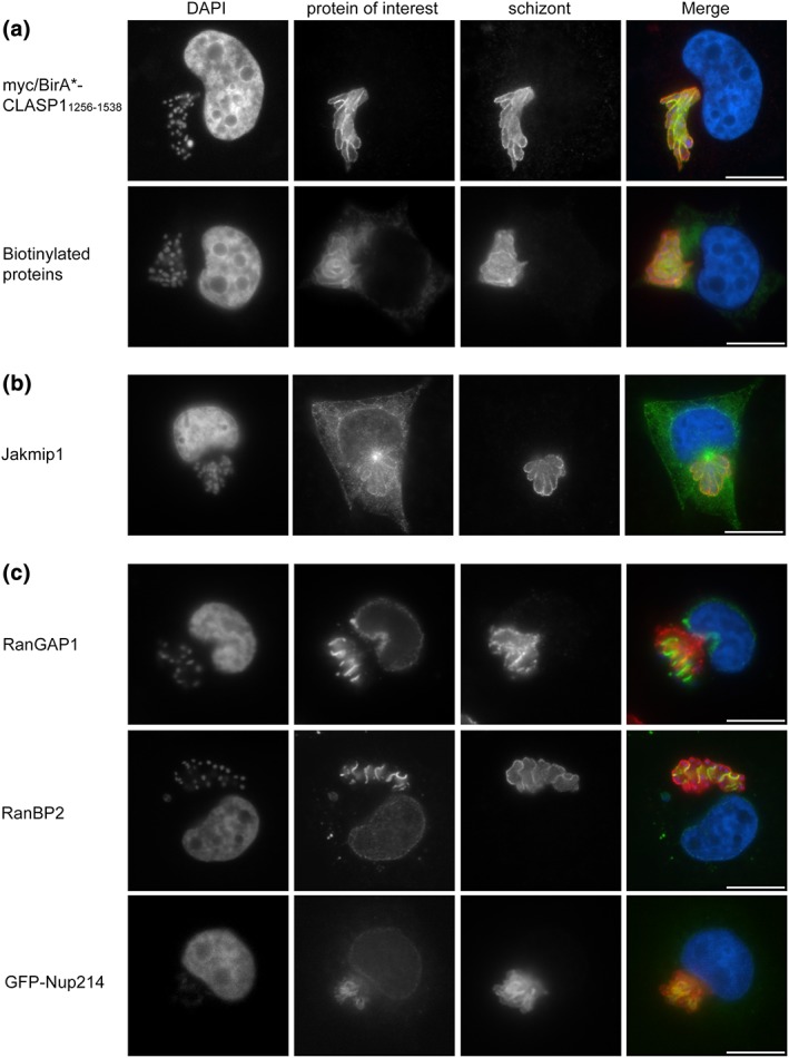
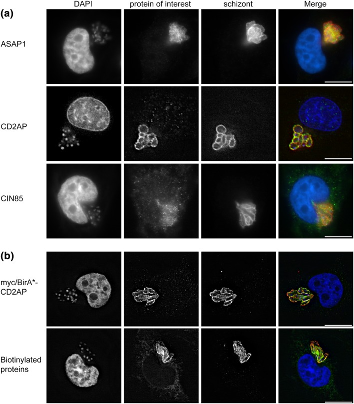
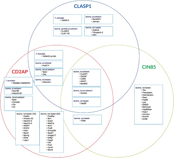
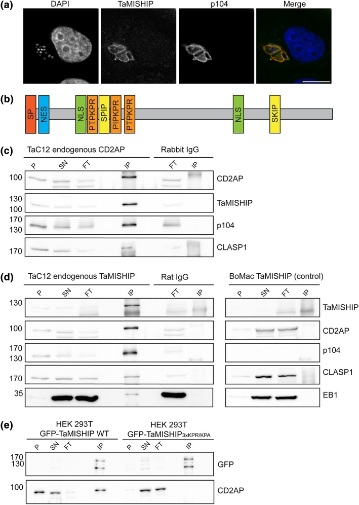
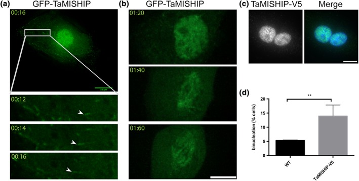
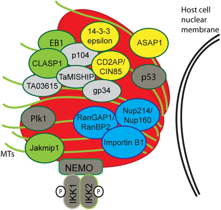
Theileria annulata is an apicomplexan parasite that modifies the phenotype of its host cell completely, inducing uncontrolled proliferation, resistance to apoptosis, and increased invasiveness. The infected cell thus resembles a cancer cell, and changes to various host cell signalling pathways accompany transformation. Most of the molecular mechanisms leading to Theileria-induced immortalization of leukocytes remain unknown. The parasite dissolves the surrounding host cell membrane soon after invasion and starts interacting with host proteins, ensuring its propagation by stably associating with the host cell microtubule network. By using BioID technology together with fluorescence microscopy and co-immunoprecipitation, we identified a CLASP1/CD2AP/EB1-containing protein complex that surrounds the schizont throughout the host cell cycle and integrates bovine adaptor proteins (CIN85, 14-3-3 epsilon, and ASAP1). This complex also includes the schizont membrane protein Ta-p104 together with a novel secreted T. annulata protein (encoded by TA20980), which we term microtubule and SH3 domain-interacting protein (TaMISHIP). TaMISHIP localises to the schizont surface and contains a functional EB1-binding SxIP motif, as well as functional SH3 domain-binding Px(P/A)xPR motifs that mediate its interaction with CD2AP. Upon overexpression in non-infected bovine macrophages, TaMISHIP causes binucleation, potentially indicative of a role in cytokinesis.

Keywords: BioID; CD2AP; Theileria; adaptor proteins; host-parasite interactions; microtubules.


Figures

Figure 1
Figure 1
Use of BioID to identify microtubule (MT)‐binding proteins and proteins of the nuclear pore complex at the schizont surface. (a) Theileria annulata‐transformed cells (TaC12) were transduced with myc‐BirA*‐CLASP11256–1538 lentivirus particles and analysed by immunofluorescence analysis. The localisation of myc‐BirA*‐CLASP11256–1538 was analysed with anti‐myc labelling (green); anti‐TaSP (red) antibodies were used to label the schizont surface (top panel). TaC12_ myc‐BirA*‐CLASP11256–1538 cells were incubated with 50‐μM biotin prior to fixation and analysis with FITC‐conjugated streptavidin (green); the parasite surface was labelled with anti‐p104 antibodies (red). DNA is labelled with DAPI (blue). (b) The MT‐interacting protein Jakmip1 associates with the parasite surface. TaC12 cells were stained with anti‐Jakmip1 (green), the parasite was labelled with anti‐p104 (red), and host and parasite nuclei were labelled with DAPI (blue). (c) Several proteins involved in nucleo‐cytoplasmic transport were identified with BioID and tested for proximity to the T. annulata schizont surface. TaC12 cells were stained with anti‐RanGAP1 (green, top panel), anti‐RanBP2 (green, middle panel), or transfected with GFP‐Nup214 (bottom panel). The parasite was labelled with anti‐p104 (red, top, and middle panels) or anti‐TaSP (red, bottom panel), and host and parasite nuclei were labelled with DAPI (blue). Scale bar = 10 μm
Figure 2
Figure 2
Analysis of bovine adaptor protein (ASAP1, CD2AP, and CIN85) localisation, and function of myc/BirA*‐CD2AP. (a) TaC12 cells were stained with anti‐ASAP1 (green, top panel), anti‐CD2AP (green, middle panel) or anti‐CIN85 (green, bottom panel). The parasite was labelled with anti‐p104 (red), and host and parasite nuclei were labelled with DAPI (blue). (b) TaC12 cells were transduced with the myc/BirA*‐CD2AP construct and analysed by immunofluorescence microscopy. Cells were stained with anti‐myc (green), and the parasite was labelled with anti‐TaSP (red); DNA was labelled with DAPI (blue, top panel). TaC12_myc‐BirA*‐CD2AP cells were incubated with 50‐μM biotin and subsequently stained with FITC conjugated streptavidin (green). The schizont was labelled with anti‐TaSP (red), and DNA was labelled with DAPI (blue, bottom panel). Scale bar = 10 μm
Figure 3
Figure 3
Venn diagram summarising the proteins identified in three BioID experiments. The proteins identified by mass spectrometry in three independent BioID experiments (myc‐BirA*‐CLASP11256–1538, myc‐BirA*‐CD2AP, and myc‐BirA*‐CIN85) are summarised in a Venn diagram. The proteins are grouped into Theileria annulata proteins and bovine proteins that localise or do not localise to the schizont, or were not tested for schizont localisation. Immunofluorescence analysis (IFA) of proteins that were found to associate with the schizont are shown in Figures 1, 2, and S1 (Jakmip1, RanGAP1, RanBP2, GFP‐Nup214, GFP‐Nup160, GFP‐Importin B1, ASAP1, CD2AP, and CIN85). The following proteins were tested in IFA, and no association with the parasite was found: GFP‐Ezrin, GFP‐Cortactin, mCherry‐Clathrin, GFP‐Talin1, CrkL‐GFP, and Coronin1B‐pmCherry. The Venn diagram was made by using the web page https://creately.com/app/#
Figure 4
Figure 4
Endogenous TaMISHIP localises to the Theileria schizont surface and interacts with CD2AP in a protein complex on the Theileria annulata schizont surface. (a) Endogenous TaMISHIP was stained with anti‐TaMISHIP (green), and the parasite was labelled anti‐p104 (red). Host and parasite nuclei were labelled with DAPI (blue). Scale bar = 10 μm. (b) TaMISHIP protein structure showing different important domains of the T. annulata protein. The protein consists of 970 amino acids and comprises two EB1‐binding SxIP motifs (yellow), several domains with nuclear localisation signals (NLS, green), a domain with a nuclear export signal (NES, blue), and SH3 domain‐binding Px(P/A)xPR motifs (orange). (c) TaC12 cells were lysed and subjected to co‐immunoprecipitation (co‐IP) with rabbit polyclonal anti‐CD2AP or rabbit IgG (control) antibodies. For each sample the nonsoluble pellet (P), lysate supernatant (SN), IP flow through (FT; for each 1.5% of total amount), and 3.3% of total bound fraction after IP (IP) was analysed by SDS‐PAGE. Analysis with anti‐CD2AP antibodies confirmed the successful immunoprecipitation of CD2AP. The co‐precipitation of the parasite proteins TaMISHIP and Ta‐p104, and the host cell protein CLASP1 could be confirmed with the corresponding antibody probes. (d) TaC12 cells were lysed and subjected to co‐immunoprecipitation (co‐IP) with rat polyclonal anti‐TaMISHIP or rat IgG (control) antibodies. As a control, the anti‐TaMISHIP immunoprecipitation was also performed with non‐infected BoMac that do not express the T. annulata protein TaMISHIP. For each sample, the nonsoluble pellet (P), lysate supernatant (SN), IP flow through (FT; for each 1.5% of total amount), and 5% of total bound fraction after IP (IP) was analysed by SDS‐PAGE. Analysis with anti‐TaMISHIP antibodies confirmed the successful immunoprecipitation of TaMISHIP. The co‐precipitation of host cell CD2AP, Ta‐p104, CLASP1, and EB1 could be confirmed with the corresponding antibody probes. (e) HEK 293T cells transfected with WT GFP‐TaMISHIP or GFP‐TaMISHIP in which all three Px(P/A)xPR motifs were mutated to Px(P/A)xPA (GFP‐TaMISHIP3XKPR➔KPA) were lysed 16 hr after transfection and subjected to co‐IP using GFP‐TRAP magnetic beads. For each sample, the nonsoluble pellet (P), lysate supernatant (SN), IP flow through (FT; for each 1% of total amount), and 10% of total bound fraction after IP (IP) was analysed by Western blotting. Analysis with anti‐eGFP (enhanced GFP) antibodies confirmed the expression of the fusion proteins and their precipitation with GFP‐TRAP beads. GFP‐TaMISHIP is predicted to run at around 136 kDa, and the additional band detected with the anti‐GFP antibody might be degradation products of the fusion protein. Although CD2AP is co‐precipitated with WT GFP‐TaMISHIP, no interaction can be seen between mutated GFP‐TaMISHIP and CD2AP.
Figure 5
Figure 5
TaMISHIP localises to the nucleus, the mitotic spindle, and MT plus ends when overexpressed in non‐infected bovine macrophages. (a) Live cell imaging was performed with BoMac cells expressing GFP‐TaMISHIP, and pictures were taken every 2 s. The top panel shows a single time point from Movie S3. The region magnified in three subsequent time points (bottom panels) is indicated with a white rectangle. MT plus‐end tracking behaviour of GFP‐TaMISHIP is indicated with arrow heads. Scale bar = 5 μm. (b) Live cell imaging was performed with BoMac cells expressing GFP‐TaMISHIP, and pictures were taken every 10 min. The top panel shows a BoMac cell just prior to mitosis, when GFP‐TaMISHIP accumulates in the nucleus. The middle and bottom panels show the progression through mitosis, and the accumulation of GFP‐TaMISHIP on the mitotic spindle. The corresponding live cell imaging can be found in Movie S4. Scale bar = 5 μm. (c) TaMISHIP‐V5 was overexpressed in BoMac cells, and fixed cells were stained with anti‐V5 (green). DNA was labelled with DAPI (blue). Scale bar = 10 μm. (d) BoMac cells were transfected with TaMISHIP‐V5 and fixed 24 hr after transfection. Binucleation was counted in nontransfected and transfected cells. Shown is the mean relative number of binucleated cells in three independent experiments (n = 131, 90, 89; p = .0031)
Figure 6
Figure 6
Graphical representation of host–parasite interactions on the Theileria annulata schizont surface. Parasite surface‐associated proteins identified in our BioID and co‐IP experiments (Ta‐p104, gp34, TaMISHIP, and TA03615) are shown in grey. Microtubule‐associating proteins found with BioID (CLASP1, Jakmip1), or previously published as binding to the parasite (EB1), are represented in green. Adaptor proteins (CD2AP, CIN85, ASAP1, and 14‐3‐3 epsilon) are represented in yellow, and proteins involved in nuclear transport (RanGAP1, RanBP2, Nup214, Nup160, and Importin B1) are represented in blue. The cell cycle‐dependent association of Plk1 with the schizont and the recruitment of the IKK signalosome complex and p53 have been described previously, and P in a circle represents phosphorylation

Similar articles

Cited by

References

    1. Adamson, R. , & Hall, R. (1997). A role for matrix metalloproteinases in the pathology and attenuation of Theileria annulata infections. Parasitol Today (Regul Ed), 13, 390–393. - PubMed
    1. Akagi, T. , Murata, K. , Shishido, T. , & Hanafusa, H. (2002). v‐Crk activates the phosphoinositide 3‐kinase/AKT pathway by utilizing focal adhesion kinase and H‐Ras. Molecular and Cellular Biology, 22, 7015–7023. - PMC - PubMed
    1. Akhmanova, A. , Hoogenraad, C. C. , Drabek, K. , Stepanova, T. , Dortland, B. , Verkerk, T. , … Galjart, N. (2001). Clasps are CLIP‐115 and ‐170 associating proteins involved in the regional regulation of microtubule dynamics in motile fibroblasts. Cell, 104, 923–935. - PubMed
    1. Bakheit, M. A. , Ahmed, J. S. , & Seitzer, U. (2008). Existence of splicing variants in homologues of Theileria lestoquardi clone‐5 gene's transcripts in Theileria annulata and Theileria parva. Annals of the New York Academy of Sciences, 1149, 212–213. - PubMed
    1. Bakheit, M. A. , Scholzen, T. , Ahmed, J. S. , & Seitzer, U. (2006). Molecular characterization of a Theileria lestoquardi gene encoding for immunogenic protein splice variants. Parasitology Research, 100, 161–170. - PubMed

Publication types

Substances