Tuberculous lumbar spinal epidural abscess in a young adult (case report)
- PMID: 29521260
- PMCID: PMC5844233
- DOI: 10.1051/sicotj/2018005
Tuberculous lumbar spinal epidural abscess in a young adult (case report)
Abstract
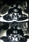
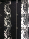
Introduction: Spinal Epidural abscess (SEA) is an uncommon pathology that needs an urgent intervention to decompress the pressure on the spinal epidural sac, cord, and roots. The authors report a rare case of a young adult with lumbar spinal epidural tuberculous abscess occupying the spinal canal from L2-L5 vertebrae with extesion to the posterior paraspinal muscles and presenting with bilateral progressive lower limb weakness. Case report: A 42 years old male teacher presented with a 15-day history of progressive difficulty to walking and bilateral lower limb weakness associated with fever, malaise and later on urinary incontinence. A magnetic resonance imaging (MRI) scan revealed a paraspinal intermuscular abscess and an abscess occupying the spinal canal compressing the dural sac from L2-L4/5, without any signs of vertebral involvement. Surgery was done by a posterior midline incision. Pus was evacuated from multiple pockets through the paraspinal muscle layers. Laminectomy for L3/4, and hemilaminectomy for L2/3, and L4/5 were performed. Pus and bone specimens were negative for acid-fast bacilli. However, both histopathological studies and Polymerase Chain Reaction (PCR) testing confirmed the presence of tuberculosis (TB). The patient received TB antibiotics, and a follow-up MRI scan at 2 months showed complete evacuation of the abscess. However, signs of L5 spondylitis were evident. No further surgery was needed as there was no vertebral collapse or neural compression and the patient's clinical condition was improving. He had normal right lower limb power and sensation and grade 4+ motor power of the left lower limb. Bowels and bladder function was normal.
Conclusion: Isolated tuberculous spinal epidural abscess is a rare disease and should be treated urgently with evacuation and decompression. Signs of spondylitis or spondylodiscitis may appear later and therefore long follow up is recommended in tuberculous cases presenting with an isolated epidural abscess.
© The Authors, published by EDP Sciences, 2018.
Figures


References
-
- Smith C, Crawford III CH, Dimar J (2014) Spinal epidural abscess: a review of diagnosis and treatment. Curr Orthop Pract 25(1): 29–33.
-
- Arora S, Kumar R (2011) Tubercular spinal epidural abscess involving the dorsal-lumbar-sacral region without osseous involvement. J Infect Dev Ctries 5(07): 544–549. - PubMed
-
- Ansari S, Rauniyar RK, Dhungel K, Sah PL, Chaudhary P, Ahmad K, et al. (2013) MR evaluation of spinal tuberculosis. Al Ameen J Med Sci 6(3): 219–225.
LinkOut - more resources
Full Text Sources
Other Literature Sources
