. 2018 Mar 9;14(3):e1007246.
doi: 10.1371/journal.pgen.1007246. eCollection 2018 Mar.

Morphogenetic defects underlie Superior Coloboma, a newly identified closure disorder of the dorsal eye

Affiliations

Morphogenetic defects underlie Superior Coloboma, a newly identified closure disorder of the dorsal eye

Jennifer C Hocking et al. PLoS Genet. .

Abstract

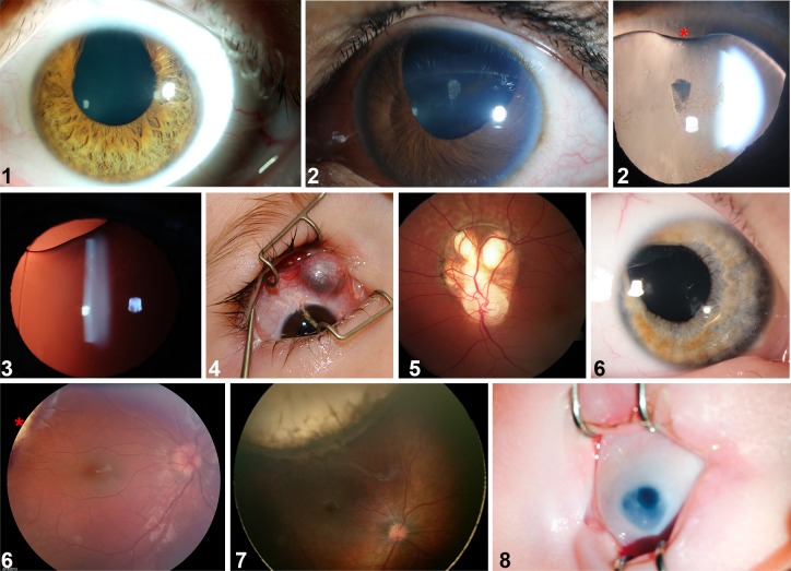
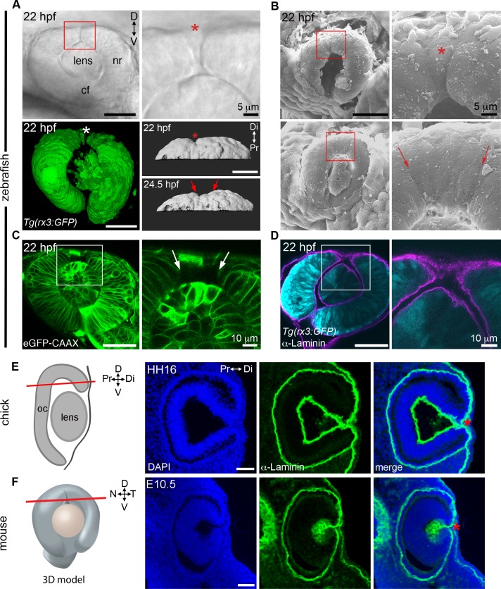
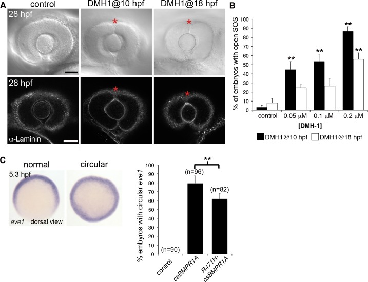
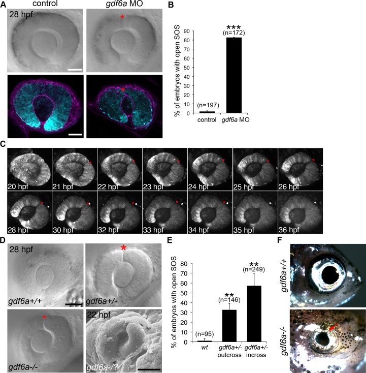
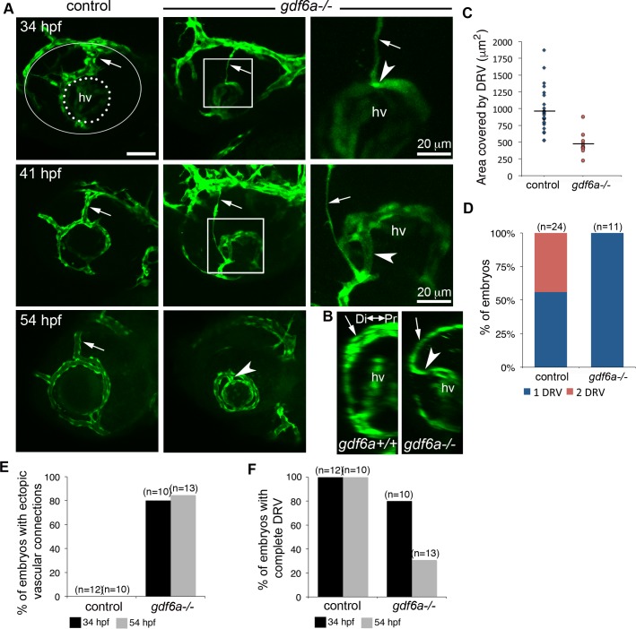
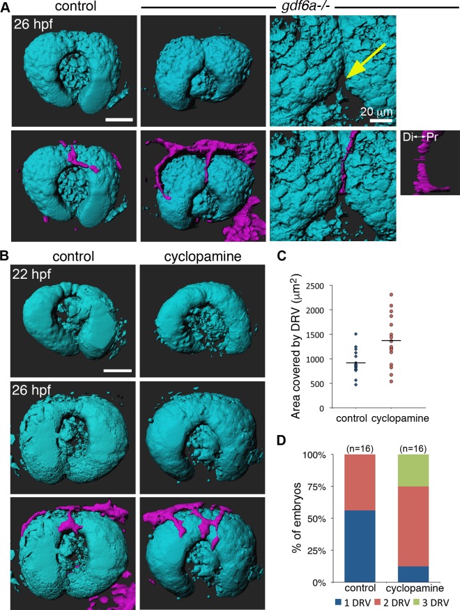
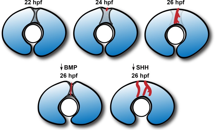
The eye primordium arises as a lateral outgrowth of the forebrain, with a transient fissure on the inferior side of the optic cup providing an entry point for developing blood vessels. Incomplete closure of the inferior ocular fissure results in coloboma, a disease characterized by gaps in the inferior eye and recognized as a significant cause of pediatric blindness. Here, we identify eight patients with defects in tissues of the superior eye, a congenital disorder that we term superior coloboma. The embryonic origin of superior coloboma could not be explained by conventional models of eye development, leading us to reanalyze morphogenesis of the dorsal eye. Our studies revealed the presence of the superior ocular sulcus (SOS), a transient division of the dorsal eye conserved across fish, chick, and mouse. Exome sequencing of superior coloboma patients identified rare variants in a Bone Morphogenetic Protein (Bmp) receptor (BMPR1A) and T-box transcription factor (TBX2). Consistent with this, we find sulcus closure defects in zebrafish lacking Bmp signaling or Tbx2b. In addition, loss of dorsal ocular Bmp is rescued by concomitant suppression of the ventral-specific Hedgehog pathway, arguing that sulcus closure is dependent on dorsal-ventral eye patterning cues. The superior ocular sulcus acts as a conduit for blood vessels, with altered sulcus closure resulting in inappropriate connections between the hyaloid and superficial vascular systems. Together, our findings explain the existence of superior coloboma, a congenital ocular anomaly resulting from aberrant morphogenesis of a developmental structure.


Conflict of interest statement

The authors have declared that no competing interests exist.

Figures

Fig 1
Fig 1. Superior coloboma.
Montage from patients with superior coloboma (numbers represent patients described in S1 Table). #1: unilateral superior iris coloboma. #2: first panel, asymmetrically-sized iris defects with bilateral pupil involvement, left eye shown; second panel, superior lenticular coloboma (asterisk) associated with a lens zonule defect. #3: lenticular coloboma, lens edge visible with retro-illumination. #4: superior scleral defect with uveal (choroid) protrusion. #5: superior retino-choroidal coloboma extending from optic disc in patient with Dandy-Walker Syndrome. #6: first panel, iris coloboma; second panel, edge of retino-chorodial coloboma (asterisk). #7: extensive retino-choroidal coloboma. #8: intra-operative photograph of a superior iris coloboma in a microphthalmic eye.
Fig 2
Fig 2. The superior ocular sulcus in zebrafish, chick and mouse.
(A) Zebrafish eyes displaying superior ocular sulci (SOS) marked by an asterisk or arrows. Top row: lateral view DIC image of the eye of a live embryo, photographed on a compound microscope. Enlarged view is shown in panel on right. Bottom row: Left, lateral view surface projection of the eye of a live Tg(rx3:GFP) embryo; Right, surface projection dorsal views of eyes from a Tg(rx3:GFP) embryo. (B) Scanning electron micrographs showing SOS at narrow (top row) and wide (bottom row) phases. Red boxes denote regions enlarged in panels on the right. (C) Single optical section, lateral view, through the eye of an embryo injected with eGFP-CAAX mRNA to label the cell membranes, with right panel showing enlarged view of boxed area. (D) Single optical section, lateral view, through eye of Tg(rx3:GFP) embryo (cyan) immunolabelled for Laminin to highlight the basal lamina (magenta). (E) Diagram showing chick eye with red line demonstrating the plane of section employed on the right. Representative horizontal section through the dorsal eye of a HH16 chick, stained with a Laminin antibody (green) and DAPI (blue). A dorsal, Laminin-lined space is evident in the distal portion of optic cup (asterisk). (F) Diagram showing 3D model of an embryonic eye with red line demonstrating plane of section for both mouse and chick sections. Right three panels are a representative horizontal section through the dorsal eye of an embryonic day 10.5 (E10.5) mouse, stained with a Laminin antibody (green) and DAPI (blue). A dorsal, Laminin-lined space is evident in the distal portion of optic cup (asterisk). Except where noted, scale bars are 50 μm. cf, choroid fissure; D-V, dorsal-ventral; HH, Hamburger Hamilton embryonic stage; hpf, hours post fertilization; N-T, nasal-temporal; nr, neural retina; Pr-Di, proximal-distal.
Fig 3
Fig 3. The role of BMPR1 signaling in closure of the superior ocular sulcus.
(A-B) Effect of Bmpr1 antagonist DMH1 on SOS closure. Lateral view DIC images of eyes from live embryos (first row) and single optical slices of eyes processed for anti-Laminin immunofluorescence (second row) following exposure to control media or 0.02 μM DMH1, starting at either 10 or 18 hpf (A). SOS is marked by red asterisk. Quantification of delayed sulcus closure in DMH1-treated embryos (B). N = 3 experiments, n = 89 or 90 embryos for each condition. Data are means ± SEM. Statistics is a one-way ANOVA for each time series with Tukey's post-hoc test: **P<0.01. (C) Injection of caBMPR1A mRNA into one-cell stage zebrafish embryos caused expansion of eve1 gene expression into a circular ring in whole embryos at 50% epiboly (5.3 hpf). Significantly fewer embryos exhibited circular eve1 expression when injected with R471H-caBMPR1A. N = 3 experiments. Data are means ± SEM. Statistics is a two-tailed t test: *P<0.05. Scale bars are 50 μm.
Fig 4
Fig 4. The role of Gdf6a signaling in superior ocular sulcus morphogenesis.
(A) Delayed SOS closure caused by Gdf6a knockdown. Tg(rx3:GFP) zebrafish eyes (cyan) from uninjected and Gdf6a morpholino-injected embryos shown as DIC images of live embryos and single optical slices following anti-Laminin antibody staining (magenta). SOS marked by red asterisk. (B) Quantification of embryos with delayed sulcus closure, as assessed at 28 hpf. (C) Time series of maximum projection confocal images of a Tg(rx3:GFP) embryo injected with gdf6a morpholino. (D) DIC images of wildtype, gdf6a+/- and gdf6a-/- eyes (SOS marked by red asterisk). Bottom right panel shows SEM image of a Gdf6a-deficient eye with a pronounced sulcus. (E) Quantification of gdf6a-/- mutants (or siblings) with delayed SOS closure. (F) Adult wildtype zebrafish (top panel) showing normal eye morphology and a gdf6a-/- zebrafish (bottom panel) with superior coloboma (red arrow). N = 3 experiments for graphs in B and E. n = number of embryos. Data are means ± SEM. Statistics in B is a two-tailed t test, and in E is one-way ANOVA with Tukey’s test: **P<0.01, *** P<0.001. Scale bars are 50 μm.
Fig 5
Fig 5. Inhibition of Hedgehog signaling rescues closure of the superior ocular sulcus in Gdf6a-deficient embryos.
(A-B) Effect of Hedgehog inhibition (cyclopamine treatment) on SOS closure in Gdf6a-deficient embryos. DIC images of gdf6a+/- eyes, treated with either control solution (left) or 10 μM cyclopamine (right) (A). SOS marked by red asterisk. Quantification of effect of cyclopamine treatment on SOS closure in gdf6a+/- incross embryos (B). (C-D) Effect of cyclopamine on dorsal retinal patterning in Gdf6a-deficient embryos. tbx5 RNA expression in eyes from 28 hpf gdf6a+/+, gdf6a+/-, and gdf6a-/- embryos with or without cyclopamine treatment (C). Quantification of effect of cyclopamine treatment on area of tbx5 expression (D). n = number of embryos, N = 4 (B) or 3 (D) experiments. Data are means ± SEM. Data in B and D are means ± SEM; Statistics in B is a one-way ANOVA with Tukey’s test, D is two-way ANOVA with Tukey’s test: **P<0.01. Scale bars are 50 μm.
Fig 6
Fig 6. Analysis of Tbx2b and closure of the superior ocular sulcus.
(A) Whole-mount in situ hybridization for zebrafish tbx2b in control and BMP-depleted embryos. Top panels are eyes dissected from control and DMH1-treated embryos; bottom panels are from gdf6a+/+, and gdf6a-/- embryos. (B-C) Analysis of SOS closure in Tbx2b-depleted embryos. DIC images of eyes from live tbx2b+/+ (top panel) and tbx2bfby (bottom panel) embryos (B). Quantification of SOS closure in wild type and tbx2bfby mutant zebrafish eyes (C). Data are means ± SEM; one-way ANOVA with Tukey’s test: *P<0.05. Scale bars are 50 μm.
Fig 7
Fig 7. Developmental functions of the superior ocular sulcus.
(A) The prominent and persistent SOS (red asterisk) present in gdf6a+/- embryos aligns with the boundary between the nasal marker foxg1a and temporal marker foxd1. Note that nasal-temporal patterning is unchanged in the gdf6a heterozygotes (bottom row) compared to the wildtype embryos (top row). (B) SEM photographs showing the dorsal radial vessel (DRV) extending into the SOS (top row). DIC images of DRV (blue arrowheads) within a wide SOS (red arrows) (bottom row). Right panels are magnified views of boxed regions. (C-D) Surface projections (C) and single optical slices (D) from confocal images of Tg(rx3:GFP; kdrl:mCherry) embryos show the DRV (magenta) extending through the SOS (optic cup and lens are cyan). Scale bars are 50 μm unless otherwise noted.
Fig 8
Fig 8. Abnormal ocular vasculature in gdf6a homozygous mutants.
(A-B) Growing blood vessels (green) in the developing eyes of gdf6a-/- or control (sibling) embryos are highlighted by the kdrl:eGFP transgene and shown as maximum projections of confocal z-stacks (A) or 90° lateral rotations thereof (B). Dorsal radial vessels (DRVs) are indicated by arrows. In the top left panel, the lens is outlined with a dotted line and the entire eye with a white line. The DRV forms in most gdf6a-/- mutants (shown at 34 hpf), can be observed degrading in 41 hpf embryos, and is often absent by 54 hpf. Ectopic connections (arrowheads) between DRV and hyaloid vasculature (hv) are visible in gdf6a-/- embryos. Right panels are enlarged views of boxed regions. (B) Laterally rotated images showing ectopic connection to hyaloid vasculature in a gdf6a-/- embryo, but not in a wildtype embryo at 41 hpf. (C-D) Quantification of area and number of DRV vessel(s) in 26 hpf control and gdf6a-/- embryos. (E-F) Quantification showing percentage of control and gdf6a-/- embryos with an ectopic connection between the hyaloid and superficial vascular systems (E) and a complete DRV (F) as assessed at 34 and 54 hpf. n = number of embryos. Scale bar is 50 μm.
Fig 9
Fig 9. Aberrant SOS closure leads to abnormal vasculature.
(A) Surface projections of 26 hpf Tg(rx3:GFP; kdrl:mCherry) wildtype and gdf6a-/- embryos, shown without vessels (top row) and with vessels (bottom row). Last column shows expanded views of same gdf6a-/- eye, highlighting the divot in the dorsal retina at the inferior edge of the superior ocular sulcus (yellow arrow). Small panel is 90° lateral rotation of vessel in adjacent panel, showing the DRV turn and extend toward the hyaloid vasculature. (B) Surface projections of Tg(rx3:GFP; kdrl:mCherry) embryos before (22 hpf) and after (26 hpf) DRV formation, with and without cyclopamine treatment. (C-D) Quantification of the area and number of DRV vessel(s) in control and cyclopamine-treated 26 hpf embryos. n = number of embryos. Scale bars are 50 μm unless otherwise noted. Di-Pr, distal-proximal.
Fig 10
Fig 10. Model of superior ocular sulcus morphogenesis and function.
The superior ocular sulcus appears as a narrow groove in the dorsal retina soon after optic cup formation (22 hpf), and subsequently becomes wider (24 hpf). The DRV grows through the wide sulcus as it travels across the dorsal retina towards the lens (24–26 hpf). If BMP signaling is reduced, the sulcus persists as a deep and narrow structure, through which the DRV still travels. However, in low BMP conditions, the DRV has a thin and unbranched morphology as it traverses the deeper fissure, and then enters the divot at the inferior edge of the sulcus and forms an ectopic connection with the hyaloid vessels. If Sonic Hedgehog signaling is reduced, the SOS is absent at the time of DRV growth, resulting in the formation of more DRV vessels spread across the dorsal retina.

Similar articles

Cited by

References

    1. Bazin-Lopez N, Valdivia LE, Wilson SW, Gestri G. Watching eyes take shape. Curr Opin Genet Dev. 2015;32: 73–79. doi: 10.1016/j.gde.2015.02.004 - DOI - PMC - PubMed
    1. Fuhrmann S. Eye morphogenesis and patterning of the optic vesicle. Curr Top Dev Biol. 2010;93: 61–84. doi: 10.1016/B978-0-12-385044-7.00003-5 - DOI - PMC - PubMed
    1. Chang L, Blain D, Bertuzzi S, Brooks BP. Uveal coloboma: clinical and basic science update. Curr Opin Ophthalmol. 2006;17: 447–470. doi: 10.1097/01.icu.0000243020.82380.f6 - DOI - PubMed
    1. Onwochei BC, Simon JW, Bateman JB, Couture KC, Mir E. Ocular colobomata. Surv Ophthalmol. 2000;45: 175–194. - PubMed
    1. Williamson KA, FitzPatrick DR. The genetic architecture of microphthalmia, anophthalmia and coloboma. Eur J Med Genet. 2014;57: 369–380. doi: 10.1016/j.ejmg.2014.05.002 - DOI - PubMed

Publication types

MeSH terms