Comparative Study
. 2018 Mar 9;13(3):e0194048.
doi: 10.1371/journal.pone.0194048. eCollection 2018.

Differential role of MyD88 and TRIF signaling in myeloid cells in the pathogenesis of autoimmune diabetes

Affiliations
Comparative Study

Differential role of MyD88 and TRIF signaling in myeloid cells in the pathogenesis of autoimmune diabetes

Ariadne Androulidaki et al. PLoS One. .

Abstract

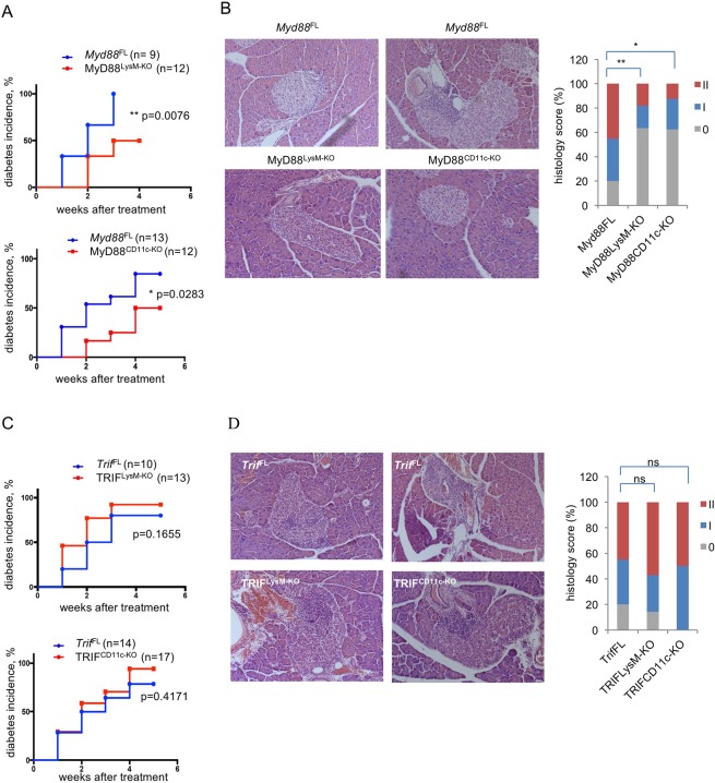
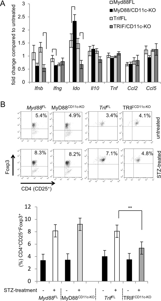
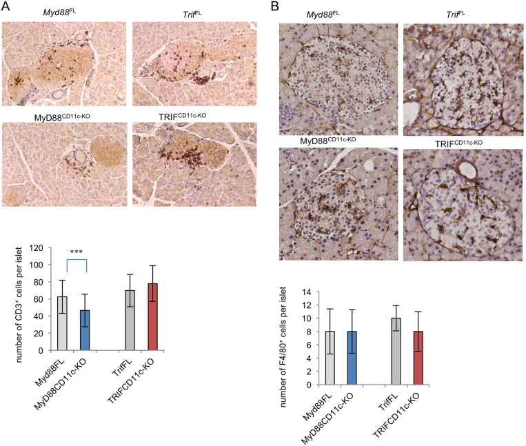
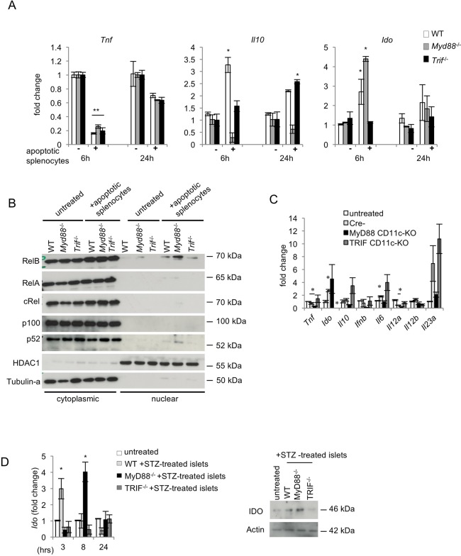
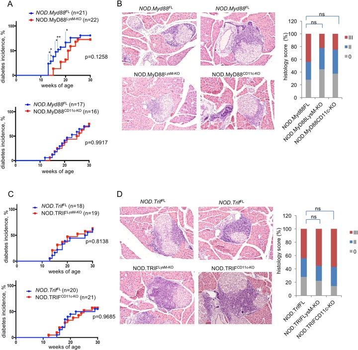
Type 1 diabetes (T1D) is caused by the autoimmune destruction of the insulin-producing pancreatic beta cells. While the role of adaptive immunity has been extensively studied, the role of innate immune responses and particularly of Toll- like Receptor (TLR) signaling in T1D remains poorly understood. Here we show that myeloid cell-specific MyD88 deficiency considerably protected mice from the development of streptozotocin (STZ)-induced diabetes. The protective effect of MyD88 deficiency correlated with increased expression of the immunoregulatory enzyme indoleamine 2,3-dioxygenase (IDO) in pancreatic lymph nodes from STZ-treated mice and in bone marrow-derived dendritic cells (BMDC) stimulated with apoptotic cells. Mice with myeloid cell specific TIR-domain-containing adapter-inducing interferon-β (TRIF) knockout showed a trend towards accelerated onset of STZ-induced diabetes, while TRIF deficiency resulted in reduced IDO expression in vivo and in vitro. Moreover, myeloid cell specific MyD88 deficiency delayed the onset of diabetes in Non-Obese Diabetic (NOD) mice, whereas TRIF deficiency had no effect. Taken together, these results identify MyD88 signaling in myeloid cells as a critical pathogenic factor in autoimmune diabetes, which is antagonized by TRIF-dependent responses. This differential function of MyD88 and TRIF depends at least in part on their opposite effects in regulating IDO expression in phagocytes exposed to apoptotic cells.


Conflict of interest statement

Competing Interests: The authors have declared that no competing interests exist.

Figures

Fig 1
Fig 1. STZ-induced diabetes development in mice with myeloid cell-specific MyD88 or TRIF deficiency.
(A and C) Graphs depicting the incidence of diabetes in mice with the indicated genotypes after treatment with 50 mg/kg STZ for five consecutive days. (B and D) Representative images and quantification of H&E stained paraffin sections of pancreatic tissue from mice with the indicated genotypes one week after completion of the STZ treatment. Graph depicts the percentage of mice with a given histology score per genotype: 0, no islet infiltrates, I, peri-insulitis, II, invasive insulitis. 30–50 islets per mouse were examined. MyD88CD11c-KO (n = 8), MyD88LysM-KO (n = 6), Myd88FL (n = 9), TRIFCD11c-KO (n = 6), TRIFLysM-KO (n = 7) and TrifFL (n = 11).
Fig 2
Fig 2. Differential effect of MyD88 and TRIF deficiency on Ido expression and Treg induction in PLNs of STZ-treated mice.
(A) The mRNA expression of the indicated genes was measured by qRT-PCR in the PLNs of MyD88CD11c-KO (n = 5), TRIFCD11c-KO (n = 9) and their respective littermate control Myd88FL (n = 4) and TrifFL (n = 8) mice one week after the completion of STZ injections. Results are depicted as fold increase compared to untreated mice (buffer-only) of each genotype. (B) FACS analysis for Tregs (CD4+CD25+Foxp3+) in the PLNs of STZ-treated mice one week after the completion of the STZ injections. Representative plots of gated live, CD4+ PLN cells stained with CD25 and intracellular Foxp3. Bar graph shows quantification of CD4+CD25+Foxp3+ cells in STZ-treated Myd88FL (n = 4), MyD88CD111c-KO (n = 6), TrifFL (n = 5) and TRIFCD11c-KO (n = 6), as well as untreated Myd88FL (n = 3), MyD88CD111c-KO (n = 2), TrifFL (n = 2) and TRIFCD11c-KO (n = 2) mice.
Fig 3
Fig 3. Immune cell infiltration in pancreatic islets of STZ-treated mice.
Paraffin sections of pancreatic islets collected from mice one week after completion of the STZ treatment were stained for CD3 (A) or F4/80 (B). CD3+ or F4/80+ cells were counted on 30–50 islets per genotype of mice scored with insulitis. Myd88FL (n = 4), MyD88CD11c-KO (n = 3), TrifFL (n = 5) and TRIFCD11c-KO (n = 6).
Fig 4
Fig 4. Differential response of MyD88- or TRIF-deficient DCs to apoptotic cell phagocytosis.
(A) WT, Myd88-/- and Trif-/- BMDCs were stimulated with apoptotic splenocytes for 6 or 24 hours and the expression of Tnf, Il10 and Ido mRNAs was measured by qRT-PCR. Data shown are representative of three independent experiments. (B) The expression of NF-κB proteins was assessed by immunoblotting with specific antibodies in cytoplasmic and nuclear extracts from BMDCs stimulated with apoptotic splenocytes for 4 hours. Tubulin and HDAC1 were used as loading controls. Blots are representative of three independent experiments. (C) The mRNA expression of the indicated genes was measured by qRT-PCR in peritoneal cells collected from MyD88CD11c-KO, TRIFCD11c-KO and Cre negative littermate control mice (n = 4 per genotype) 18 hours after i.p. injection of 107 apoptotic thymocytes or medium-only. Data are representative of two independent experiments. (D) The mRNA and protein expression of IDO was analyzed by qRT-PCR and immunoblotting respectively in WT, Myd88-/- and Trif-/- BMDCs that were stimulated with primary apoptotic islets (immunoblot was performed on protein extracts prepared from islets stimulated for 12 hours with STZ). Data are representative of two independent experiments.
Fig 5
Fig 5. Development of autoimmune diabetes in NOD mice with myeloid- specific MyD88 or TRIF deficiency.
(A and C) Graphs depicting the incidence of diabetes in mice with the indicated genotypes. (B and D) Representative images and quantification of H&E stained paraffin sections of pancreatic tissue from 10-week-old mice with the indicated genotypes. Graph depicts the percentage of mice with a given histology score per genotype: 0 or I, no islet infiltrates or only small peri-islet infiltrates; II, invasive insulitis (<50% of islet area); III, severe invasive insulitis (>50% of islet area). 20–30 islets per mouse were counted. NOD.MyD88CD11c-KO (n = 8), NOD.MyD88LysM-KO (n = 9), NOD.TRIFCD11c-KO (n = 7) and NOD.TRIFLysM-KO (n = 9), NOD.Myd88FL (n = 13), and NOD.TrifFL (n = 12).

References

    1. Lehuen A, Diana J, Zaccone P, Cooke A. Immune cell crosstalk in type 1 diabetes. Nat Rev Immunol. 2010;10(7):501–13. Epub 2010/06/26. doi: 10.1038/nri2787. . - DOI - PubMed
    1. Anderson MS, Bluestone JA. The NOD mouse: a model of immune dysregulation. Annu Rev Immunol. 2005;23:447–85. Epub 2005/03/18. doi: 10.1146/annurev.immunol.23.021704.115643. . - DOI - PubMed
    1. Pearson JA, Wong FS, Wen L. The importance of the Non Obese Diabetic (NOD) mouse model in autoimmune diabetes. J Autoimmun. 2016;66:76–88. Epub 2015/09/26. doi: 10.1016/j.jaut.2015.08.019. ; PubMed Central PMCID: PMCPMC4765310. - DOI - PMC - PubMed
    1. Lenzen S. The mechanisms of alloxan- and streptozotocin-induced diabetes. Diabetologia. 2008;51(2):216–26. Epub 2007/12/19. doi: 10.1007/s00125-007-0886-7. . - DOI - PubMed
    1. Paik SG, Fleischer N, Shin SI. Insulin-dependent diabetes mellitus induced by subdiabetogenic doses of streptozotocin: obligatory role of cell-mediated autoimmune processes. Proc Natl Acad Sci U S A. 1980;77(10):6129–33. Epub 1980/10/01. ; PubMed Central PMCID: PMCPMC350227. - PMC - PubMed

Publication types

MeSH terms

Substances