. 2018 Dec 1;444 Suppl 1(Suppl 1):S202-S208.
doi: 10.1016/j.ydbio.2018.02.020. Epub 2018 Mar 6.

Characterization of Pax3 and Sox10 transgenic Xenopus laevis embryos as tools to study neural crest development

Affiliations

Characterization of Pax3 and Sox10 transgenic Xenopus laevis embryos as tools to study neural crest development

Mansour Alkobtawi et al. Dev Biol. .

Abstract

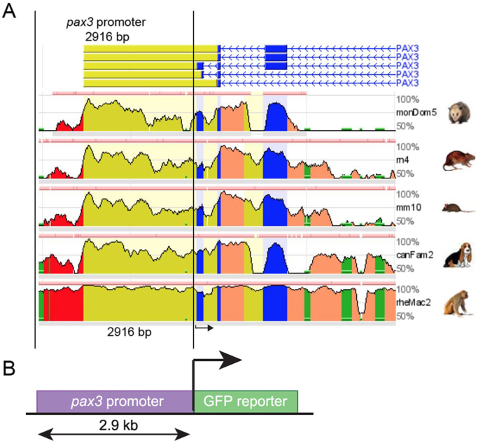
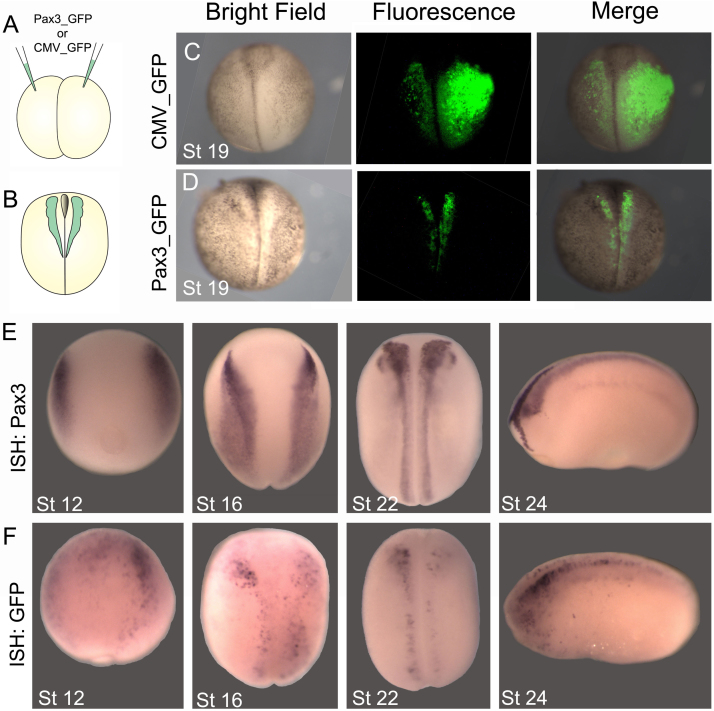
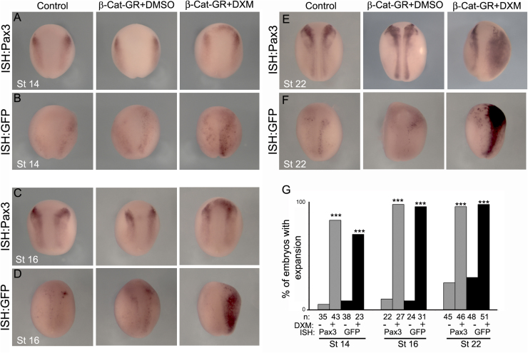
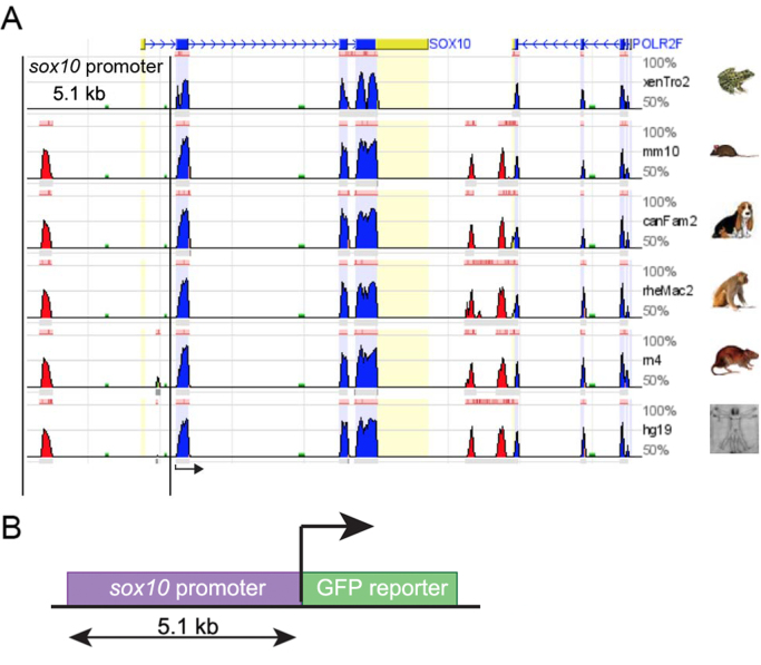
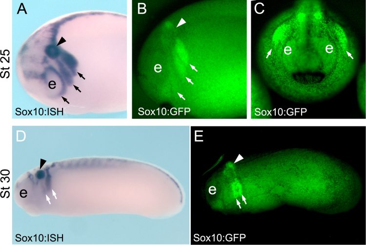
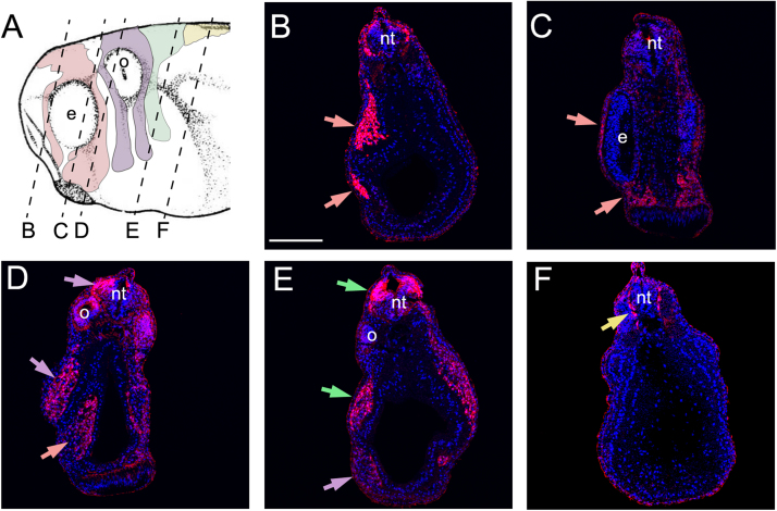
The neural crest is a multipotent population of cells that originates a variety of cell types. Many animal models are used to study neural crest induction, migration and differentiation, with amphibians and birds being the most widely used systems. A major technological advance to study neural crest development in mouse, chick and zebrafish has been the generation of transgenic animals in which neural crest specific enhancers/promoters drive the expression of either fluorescent proteins for use as lineage tracers, or modified genes for use in functional studies. Unfortunately, no such transgenic animals currently exist for the amphibians Xenopus laevis and tropicalis, key model systems for studying neural crest development. Here we describe the generation and characterization of two transgenic Xenopus laevis lines, Pax3-GFP and Sox10-GFP, in which GFP is expressed in the pre-migratory and migratory neural crest, respectively. We show that Pax3-GFP could be a powerful tool to study neural crest induction, whereas Sox10-GFP could be used in the study of neural crest migration in living embryos.

Keywords: Neural crest; Pax3; Sox10; induction; migration; transgenic.


Figures

Fig. 1
Fig. 1
Characterization of apax3promoter. (A) Graphical representation showing the result of a comparative genomic analysis obtained using the ECR browser. The genomic region corresponding to the vertebrate pax3 gene was compared among: opossum, rat, mouse, dog, and macaque. The pax3 promoter region (2916 bp) is delimited by two black parallel lines. Black arrow indicates the initiation site. bp, base-pairs. (B) Schematic showing a simplified view of the transgene containing the pax3 promoter (2916 bp) driving expression of the green-fluorescent-protein (GFP). Key: red, highly conserved elements; blue, coding exons; yellow, UTRs; green, transposable elements and simple repeats.
Fig. 2
Fig. 2
Generation of Pax3-GFP transient transgenic embryos. (A) pax3 or CMV promoters fused to GFP were injected into Xenopus embryos as described to generate transient transgenic embryos. (B) GFP fluorescence was examined at neurula stage (stage 19). (C) CMV-GFP transient transgenic embryo, showing ubiquitous expression. (D) Pax3-GFP transient transgenic embryo showing expression restricted to the neural folds. (E, F) In situ hybridization (ISH) for comparison of endogenous Pax3 expression with GFP expression in Pax3-GFP transient transgenic embryos. (E) ISH for pax3 at the indicated stages. (F) ISH for GFP in Pax3-GFP embryos at the indicated stages. Note the similar expression of Pax3 and GFP. 92% of Pax3-GFP embryos exhibited expression in the neural folds (n= 210).
Fig. 3
Fig. 3
Pax3-GFP responds to Wnt signaling. (A, C, E) ISH for endogenous Pax3 in wild type embryos at the indicated stages. (B, D, F) ISH for GFP in Pax3-GFP transient transgenic embryos at the indicated stages. Embryos were non-injected (Control) or injected with an inducible form of β-catenin (β-Cat-GR) and treated with DXM (in DMSO) to induce β-catenin, or DMSO alone. (G) Quantification of the percentage of embryos that showed expansion in Pax3/GFP expression. Note that activation of β-catenin leads to an equivalent increase of Pax3 and GFP expression. *** P<0.001.
Fig. 4
Fig. 4
Characterization ofsox10promoter. (A) Graphical representation showing the result of a comparative genomic analysis obtained using the ECR browser. The genomic region corresponding to the vertebrate sox10 gene was compared among: Xenopus tropicalis, mouse, dog, macaque, rat, and human. The sox10 promoter region (5.1 kb) is delimited by two black parallel lines. (B) Schematic showing a simplified view of the transgene containing the sox10 promoter (5.1 kb) driving the expression of the green-fluorescent-protein (GFP). Key: red, highly conserved elements; blue, coding exons; yellow, UTRs; green, transposable elements and simple repeats. Black arrow indicates the initiation site. bp, base-pairs.
Fig. 5
Fig. 5
Characterization of Sox10-GFP stable transgenic embryos. (A-C) Stage 25 embryos. (A) ISH for endogenous sox10 in wild-type embryos at the indicated stage, side view. (B, C) GFP fluorescence of Sox10-GFP stable transgenic embryos at the indicated stage, B, side view and C, anterior view. (D, E) Stage 30 embryos, side view. (D) ISH for endogenous sox10 in wild type embryos at the indicated stage. (E) GFP fluorescence of Sox10-GFP stable transgenic embryos at the indicated stage. Note the similar expression of Sox10 and GFP in the migrating neural crest cells (arrows). Arrow head indicates the otic vesicle and e the eye.
Fig. 6
Fig. 6
GFP expression in migrating neural crest in Sox10-GFP stable transgenic embryos. (A) Diagram indicating the level of the transverse sections shown in panels B-F. Pink shows mandibular neural crest; Purple shows hyoid neural crest; Green shows branchial neural crest; Yellow shows trunk/vagal neural crest. (B-F) Sections of Sox10-GFP stable transgenic embryos after immunostaining for GFP and DAPI. (B, C) Mandibular neural crest. (D) Hyoid and mandibular neural crest. (E) Branchial and hyoid neural crest. (F) Trunk/vagal neural crest. e: eye; o- otic vesicle; nt: neural tube. Epidermal immunostaining is likely to be non-specific background.

References

    1. Aoki Y., Saint-Germain N., Gyda M., Magner-Fink E., Lee Y.H., Credidio C., Saint-Jeannet J.P. Sox10 regulates the development of neural crest-derived melanocytes in Xenopus. Dev Biol. 2003;259:19–33. - PubMed
    1. Bang A.G., Papalopulu N., Kintner C., Goulding M.D. Expression of Pax-3 is initiated in the early neural plate by posteriorizing signals produced by the organizer and by posterior non-axial mesoderm. Development. 1997;124:2075–2078. - PubMed
    1. Bronner M.E., Simões-Costa M. The Neural Crest Migrating into the Twenty-First Century. Curr Top Dev Biol. 2016;116:115–134. - PMC - PubMed
    1. Corpening J.C.1, Deal K.K., Cantrell V.A., Skelton S.B., Buehler D.P., Southard-Smith E.M. Isolation and live imaging of enteric progenitors based on Sox10-Histone2BVenus transgene expression. Genesis. 2011;49:599–618. - PMC - PubMed
    1. Domingos P.M., Itasaki N., Jones C.M., Mercurio S., Sargent M.G., Smith J.C., Krumlauf R. The Wnt/beta-catenin pathway posteriorizes neural tissue in Xenopus by an indirect mechanism requiring FGF signalling. Dev Biol. 2001;239:148–160. - PubMed

Publication types

MeSH terms

Substances

LinkOut - more resources