Review
. 2018 Mar 8;172(6):1271-1293.
doi: 10.1016/j.cell.2018.01.014.

Subcellular Organization: A Critical Feature of Bacterial Cell Replication

Affiliations
Review

Subcellular Organization: A Critical Feature of Bacterial Cell Replication

Ivan V Surovtsev et al. Cell. .

Abstract

Spatial organization is a hallmark of all living systems. Even bacteria, the smallest forms of cellular life, display defined shapes and complex internal organization, showcasing a highly structured genome, cytoskeletal filaments, localized scaffolding structures, dynamic spatial patterns, active transport, and occasionally, intracellular organelles. Spatial order is required for faithful and efficient cellular replication and offers a powerful means for the development of unique biological properties. Here, we discuss organizational features of bacterial cells and highlight how bacteria have evolved diverse spatial mechanisms to overcome challenges cells face as self-replicating entities.


Figures

Figure 1
Figure 1. Shaping the cell
A. (i) FtsZ-dependent inward growth of PG during division produces new cell halves in spherical bacteria. Two types of cell cycle are shown. The top panel showcases septal PG synthesis followed by an abrupt separation of daughter cells, as in Staphylococcus aureus. The bottom panel illustrates the hypothetical case of septal PG synthesis occurring concurrently with cell constriction. A good candidate for this cell cycle pattern is Moraxella catarrhalis (Zhou et al, 2016). (ii) Septal PG growth gives rise to cell poles in rod-shaped bacteria. B. Rod shapes can be created by MreB-dependent (i) lateral or (ii) zonal PG incorporation. Preferential localization of MreB to the inner curve of a bent cell cylinder has been proposed to help restore straight shape. (iii) Rod shape can also derive from polar growth, which, in some bacteria, is DivIVA-dependent. C. Curved cell shapes can be achieved (i) by a filamentous structure such as periplasmic flagella applying an external force on the PG, (ii) through asymmetric PG growth between lateral walls caused by a static protein filament, (iii) via local modifications of the PG mesh, affecting its material properties. D. Stalk formation is achieved through localized PG growth with the assistance of a bactofilin structure.
Figure 2
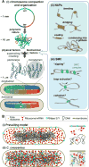
Figure 2. Folding and decoding the genome
A. (i) Bacterial chromosomes can fold into two major configurations: longitudinal or transverse. Folding is driven by physical factors and DNA-binding proteins such as (ii) NAPs and (iii) SMC complexes. SMC complexes may travel in pairs (Badrinarayanan et al., 2012; Wang et al., 2017), but just a single complex is shown for simplicity. B. (i) Prevailing view of nucleoid (transcription), polysome (translation) and RNase E or Y (mRNA degradation) arrangement in bacterial cells. (ii) Unconventional organization of the chromosomal DNA, ribosomes and RNase E in C. crescentus.
Figure 3
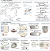
Figure 3. Partitioning cellular components
A. Different approaches ensure faithful partitioning of low-copy-number cellular components between daughter cells: (i) Cytoskeleton-based active transport of component copies to opposite poles, as illustrated with the plasmid ParM/R system. (ii) Localization of copies at opposite poles. (iii) Regular distribution of copies along the long cell axis. B. (i) Current models of the ParA/B DNA partitioning system propose different sources of the translocating force, which, together with the ParA/B biochemistry, drive (ii) oscillations of a single plasmid, (iii) regular patterning of multiple plasmid copies, and (iv) segregation of chromosomal ori regions in some bacteria. C. Segregation of chromosomal ter regions across the septal plane is driven by the hexameric FtsK translocase, which interacts with the division machinery. FstK recognizes DNA sequences on the chromosome that have an ori-to-ter orientation, which determines the directionality of DNA translocation across the septal plane.
Figure 4
Figure 4. Selecting the division site
A. Nucleoid occlusion of Z-ring formation is facilitated by the asymmetry in DNA-binding sites of FtsZ inhibitors SlmA (E. coli) or Noc (B. subtilis) along the chromosome. B. The E. coli Min system prevents Z-ring formation in the polar regions through protein oscillations driven by a reaction-diffusion mechanism. These oscillations result in time-averaged accumulation of an FtsZ-assembly inhibitor at the polar regions. C. The Pom system in M. xanthus identifies midcell by positioning an FtsZ assembly activating complex, the PomX/Y cluster, at mid-nucleoid through a localized-sink/distributed-source mechanism coupled to a translocating force of chemophoretic or chromosomal origin.
Figure 5
Figure 5. Specializing the cell
A. Protein localization at a specific cell pole results in cellular asymmetry that underlies (i) the coordination between developmental and cell cycle events in C. crescentus (the polar localization of only two histidine kinases, DivJ and PleC, are shown for clarity (Wheeler and Shapiro, 1999); in reality, there are at least a dozen regulators that display polar accumulation during the cell cycle), (ii) the actin-based motility of some intracellular pathogens and their cell-to-cell spread in eukaryotic host tissues, (iii) the directional motility in M. xanthus. B. Pole recognition can be based on (i) differences in lipid composition between membrane regions of different curvatures, (ii) sensing of high curvature by individual protein oligomers, (iii) sensing of moderate curvature through collective oligomer interactions, (iv) interplay between protein cluster diffusion and protein cluster agglomeration, (v) volume exclusion that promotes formation of large protein structures outside the nucleoid. C. Examples of bacterial microcompartments: (i) chlorosomes bound by a single lipid layer and a protein baseplate, (ii) an anammoxosome enclosed by a lipid bilayer, (iii) iron-containing magnetosomes, (iv) carboxysomes, which confine reactions involved in carbon fixation in a protein shell, (v) an endospore maturing into a spore inside the mother cell. B. subtilis SpoVM localizes to the outer surface of the endospore to facilitate spore coat formation.

Similar articles

Cited by

References

    1. Aaron M, Charbon G, Lam H, Schwarz H, Vollmer W, Jacobs-Wagner C. The tubulin homologue FtsZ contributes to cell elongation by guiding cell wall precursor synthesis in Caulobacter crescentus. Mol Microbiol. 2007;64:938–52. - PubMed
    1. Abdul-Rahman F, Petit E, Blanchard JL. Distribution of polyhedral bacterial microcompartments suggests frequent horizontal transfer and operon reassembly. J Phylogen Evolution Biol. 2013;1:118.
    1. Adachi S, Hori K, Hiraga S. Subcellular positioning of F plasmid mediated by dynamic localization of SopA and SopB. J Mol Biol. 2006;356:850–63. - PubMed
    1. Adams DW, Wu LJ, Errington J. Nucleoid occlusion protein Noc recruits DNA to the bacterial cell membrane. EMBO J. 2015;34:491–501. - PMC - PubMed
    1. Ah-Seng Y, Rech J, Lane D, Bouet JY. Defining the role of ATP hydrolysis in mitotic segregation of bacterial plasmids. PLoS Genet. 2013;9:e1003956. - PMC - PubMed

Publication types

MeSH terms

LinkOut - more resources