

Changes in MLST profiles and biotypes of Corynebacterium diphtheriae isolates from the diphtheria outbreak period to the period of invasive infections caused by nontoxigenic strains in Poland (1950-2016)
- PMID: 29523087
- PMCID: PMC5845185
- DOI: 10.1186/s12879-018-3020-1
Changes in MLST profiles and biotypes of Corynebacterium diphtheriae isolates from the diphtheria outbreak period to the period of invasive infections caused by nontoxigenic strains in Poland (1950-2016)
Abstract
Background: Corynebacterium diphtheriae is a re-emerging pathogen in Europe causing invasive infections in vaccinated persons and classical diphtheria in unvaccinated persons. In the presented study we analysed genetic changes in C. diphtheriae isolates collected in Poland from the period before the introduction of the mass anti-diphtheria vaccination to the present time when over 98% of the population is vaccinated.
Methods: A total of 62 C. diphtheriae isolates collected in the 1950s-1960s, 1990s and 2000-2016 in Poland were investigated. Examined properties of the isolates included toxigenic status, presence of tox gene, biotype, MLST type (ST) and type of infection.
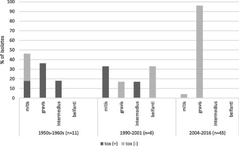
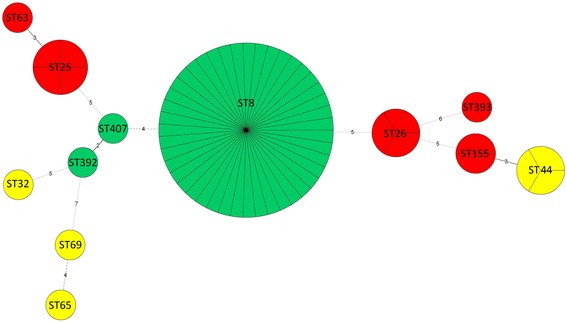
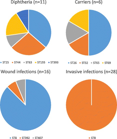
Results: A total of 12 sequence types (STs) were identified among the analysed C. diphtheriae isolates. The highest variability of STs was observed among isolates from diphtheria and asymptomatic carriers collected in the XX century. Over 95% of isolates collected from invasive and wound infections in 2004-2016 belonged to ST8. Isolates from the XX century represented all four biotypes: mitis, gravis, intermedius and belfanti, but the belfanti biotype appeared only after the epidemic in the 1990s. All except three isolates from the XXI century represented the biotype gravis.
Conclusions: During a diphtheria epidemic period, non-epidemic clones of C. diphtheriae might also disseminate and persist in a particular area after the epidemic. An increase of the anti-diphtheria antibody level in the population causes not only the elimination of toxigenic strains from the population but may also influence the reduction of diversity of C. diphtheriae isolates. MLST types do not reflect the virulence of isolates. Each ST can be represented by various virulent variants representing various pathogenic capacities, for example toxigenic non-invasive, nontoxigenic invasive and nontoxigenic non-invasive.
Keywords: Corynebacterium diphtheriae; Diphtheria; Invasive infections; MLST.
Conflict of interest statement
Ethics approval and consent to participate
Ethical approval was not required for the use of clinical samples because all the investigated isolates were taken as a part of standard care (diagnostic purposes). The samples were not collected for research purposes.
Consent for publication
Not applicable.
Competing interests
The authors declare that they have no competing interests.
Publisher’s Note
Springer Nature remains neutral with regard to jurisdictional claims in published maps and institutional affiliations.
Figures
References
-
- Zasada AA. Corynebacterium diphtheriae infections currently and in the past. Przegl Epidemiol. 2015;69:439–444. - PubMed
-
- De Winter LM, Bernard KA, Romney MC. Human clinical isolates of Corynebacterium diphtheriae and Corynebacterium ulcerans collected in Kanada form 1999 to 2003 but not fitting reporting criteria for cases of diphtheria. J Clin Microbiol. 2005;43:3447–3449. doi: 10.1128/JCM.43.7.3447-3449.2005. - DOI - PMC - PubMed
Publication types
MeSH terms
Grants and funding
LinkOut - more resources
Full Text Sources
Other Literature Sources
Medical
