

Comparison of mesenchymal stem cells obtained by suspended culture of synovium from patients with rheumatoid arthritis and osteoarthritis
- PMID: 29523119
- PMCID: PMC5845365
- DOI: 10.1186/s12891-018-1998-6
Comparison of mesenchymal stem cells obtained by suspended culture of synovium from patients with rheumatoid arthritis and osteoarthritis
Abstract
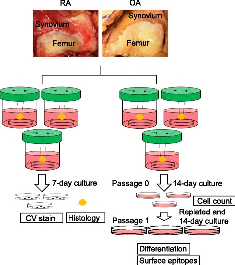
Background: Mobilization of mesenchymal stem cells (MSCs) from the synovium was revealed using a "suspended synovium culture model" of osteoarthritis (OA). The pathology of rheumatoid arthritis (RA) differs from that of OA. We investigated whether mobilization of MSCs from the synovium also occurred in RA, and we compared the properties of synovial MSCs collected from suspended synovium culture models of RA and OA.
Methods: Human synovium was harvested during total knee arthroplasty from the knee joints of patients with RA (n = 8) and OA (n = 6). The synovium was suspended in a bottle containing culture medium and a culture dish at the bottom. Cells were harvested from the dish and analyzed.
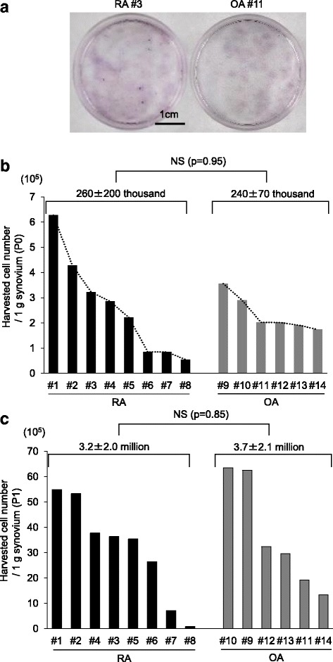
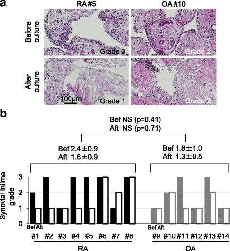
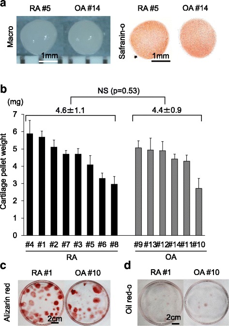
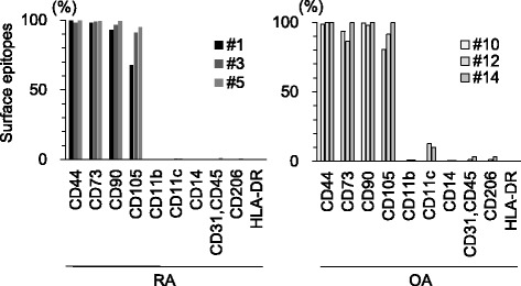
Results: No significant difference was observed between RA and OA in the harvested cell numbers per g of synovium. However, the variation in the number of cells harvested from each donor was greater for RA than for OA. The harvested cells were multipotent and no difference was observed in the cartilage pellet weight between RA and OA. The surface epitopes of the cells in RA and OA were similar to those of MSCs.
Conclusion: Mobilization of MSCs from the synovium was demonstrated using a suspended synovium culture model for RA. The harvested cell numbers, chondrogenic potentials, and surface epitope profiles were comparable between the RA and OA models.
Keywords: Chondrogenic potential; Harvested cell number; MSC mobilization; Osteoarthritis; Rheumatoid arthritis; Suspended synovium culture model; Synovial mesenchymal stem cell (synovial MSC).
Conflict of interest statement
Ethics approval and consent to participate
This study was approved by local institutional review boards (the Medical Research Ethics Committee of Tokyo Medical and Dental University and the Hospital Ethics Committee of Juntendo University Hospital), and written consent was obtained from all study subjects.
Consent for publication
Not applicable.
Competing interests
The authors declare that they have no competing interests.
Publisher’s Note
Springer Nature remains neutral with regard to jurisdictional claims in published maps and institutional affiliations.
Figures
Similar articles
-
Yields and chondrogenic potential of primary synovial mesenchymal stem cells are comparable between rheumatoid arthritis and osteoarthritis patients.Stem Cell Res Ther. 2017 May 16;8(1):115. doi: 10.1186/s13287-017-0572-8. Stem Cell Res Ther. 2017. PMID: 28511664 Free PMC article.
-
Fibrous Synovium Releases Higher Numbers of Mesenchymal Stem Cells Than Adipose Synovium in a Suspended Synovium Culture Model.Arthroscopy. 2017 Apr;33(4):800-810. doi: 10.1016/j.arthro.2016.09.033. Epub 2016 Dec 30. Arthroscopy. 2017. PMID: 28043752
-
Yields of mesenchymal stromal cells from synovial fluid reflect those from synovium in patients with rheumatoid arthritis.Tissue Cell. 2022 Apr;75:101727. doi: 10.1016/j.tice.2021.101727. Epub 2022 Jan 4. Tissue Cell. 2022. PMID: 34998163
-
Bone marrow mesenchymal stem cells in rheumatoid arthritis, spondyloarthritis, and ankylosing spondylitis: problems rather than solutions?Arthritis Res Ther. 2019 Nov 13;21(1):239. doi: 10.1186/s13075-019-2014-8. Arthritis Res Ther. 2019. PMID: 31722720 Free PMC article. Review.
-
Tenascin-C in Osteoarthritis and Rheumatoid Arthritis.Front Immunol. 2020 Sep 30;11:577015. doi: 10.3389/fimmu.2020.577015. eCollection 2020. Front Immunol. 2020. PMID: 33101302 Free PMC article. Review.
Cited by
-
Gene Expression Signatures of Synovial Fluid Multipotent Stromal Cells in Advanced Knee Osteoarthritis and Following Knee Joint Distraction.Front Bioeng Biotechnol. 2020 Oct 14;8:579751. doi: 10.3389/fbioe.2020.579751. eCollection 2020. Front Bioeng Biotechnol. 2020. PMID: 33178674 Free PMC article.
-
Comparison of the short-term effect of intra-articular hyaluronic acid and platelet-rich plasma injections in knee osteoarthritis: a randomized clinical trial.J Prev Med Hyg. 2024 Aug 31;65(2):E214-E220. doi: 10.15167/2421-4248/jpmh2024.65.2.3270. eCollection 2024 Jun. J Prev Med Hyg. 2024. PMID: 39430992 Free PMC article. Clinical Trial.
-
Cryopreservation in 95% serum with 5% DMSO maintains colony formation and chondrogenic abilities in human synovial mesenchymal stem cells.BMC Musculoskelet Disord. 2019 Jul 6;20(1):316. doi: 10.1186/s12891-019-2700-3. BMC Musculoskelet Disord. 2019. PMID: 31279341 Free PMC article.
-
Device-Based Enrichment of Knee Joint Synovial Cells to Drive MSC Chondrogenesis Without Prior Culture Expansion In Vitro: A Step Closer to 1-Stage Orthopaedic Procedures.Am J Sports Med. 2022 Jan;50(1):152-161. doi: 10.1177/03635465211055164. Epub 2021 Nov 15. Am J Sports Med. 2022. PMID: 34779670 Free PMC article.
-
Optimal Pore Size of Honeycomb Polylactic Acid Films for In Vitro Cartilage Formation by Synovial Mesenchymal Stem Cells.Stem Cells Int. 2021 Aug 2;2021:9239728. doi: 10.1155/2021/9239728. eCollection 2021. Stem Cells Int. 2021. PMID: 34394358 Free PMC article.
References
Publication types
MeSH terms
Grants and funding
LinkOut - more resources
Full Text Sources
Other Literature Sources
Medical
