Lipid accelerating the fibril of islet amyloid polypeptide aggravated the pancreatic islet injury in vitro and in vivo
- PMID: 29523142
- PMCID: PMC5845206
- DOI: 10.1186/s12944-018-0694-8
Lipid accelerating the fibril of islet amyloid polypeptide aggravated the pancreatic islet injury in vitro and in vivo
Abstract
Background: The fibrillation of islet amyloid polypeptide (IAPP) triggered the amyloid deposition, then enhanced the loss of the pancreatic islet mass. However, it is not clear what factor is the determinant in development of the fibril formation. The aim of this study is to investigate the effects of lipid on IAPP fibril and its injury on pancreatic islet.
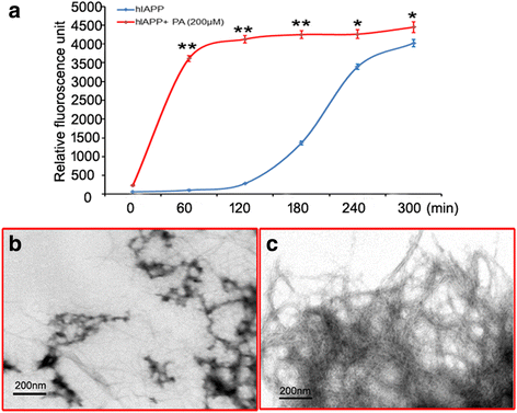
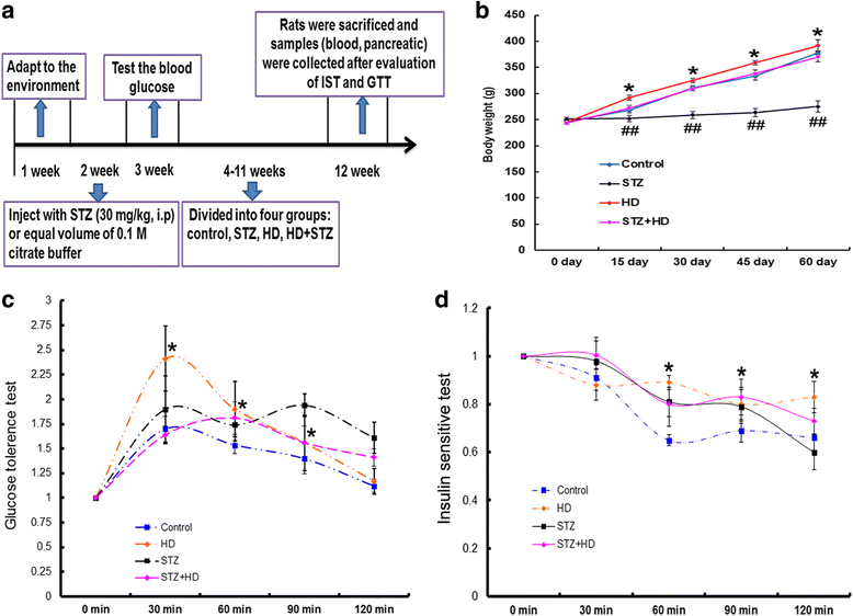
Methods: The fibril form of human IAPP (hIAPP) was tested using thioflavin-T fluorescence assay and transmission electron microscope technology after incubated with palmitate for 5 h at 25 °C. The cytotoxicity of fibril hIAPP was evaluated in INS-1 cells through analyzing the leakage of cell membrane and cell apoptosis. Type 2 diabetes mellitus (T2DM) animal model was induced with low dose streptozotocin combined the high-fat diet feeding for two months in rats. Plasma biochemistry parameters were measured before sacrificed. Pancreatic islet was isolated to evaluate their function.
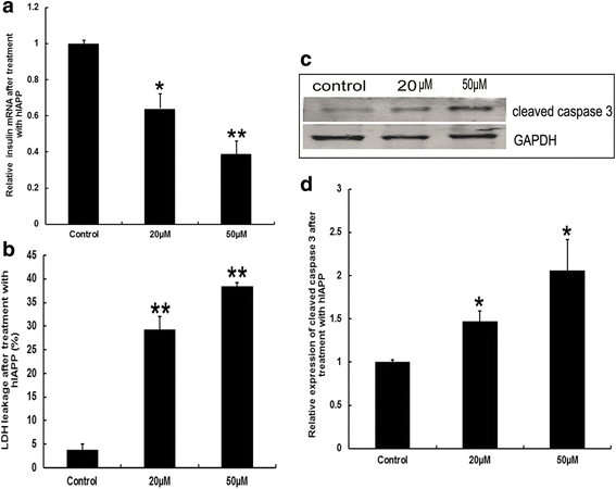
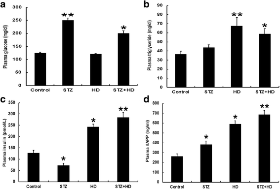
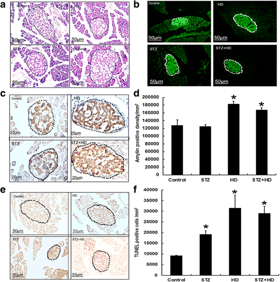
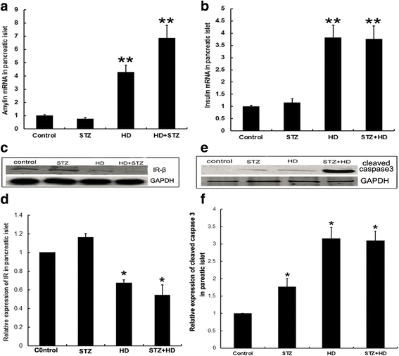
Results: The results showed that co-incubation of hIAPP and palmitate induced more fibril form. Fibril hIAPP induced cell lesions including cell membrane leakage and cell apoptosis accompanied insulin mRNA decrease in INS-1 cell lines. In vivo, Plasma glucose, triglyceride, rIAPP and insulin increased in T2DM rats compared with the control group. In addition, IAPP and insulin mRNA increased in pancreatic islet of T2DM rats. Furthermore, T2DM induced the reduction of insulin receptor expression and cleaved caspase-3 overexpression in pancreatic islet.
Conclusions: Results in vivo and in vitro suggested that lipid and IAPP plays a synergistic effect on pancreatic islet cell damage, which implicated in enhancing the IAPP expression and accelerating the fibril formation of IAPP.
Keywords: IAPP; Insulin; Lipid; Palmitate; Pancreatic islet; T2DM.
Conflict of interest statement
Ethics approval
This animal study was approved by the institutional Animal Ethics Committee, Lanzhou University (permit number: SCXK Gan 2009–0004).
Consent for publication
Not applicable
Competing interests
The authors declare that they have no competing interests.
Publisher’s Note
Springer Nature remains neutral with regard to jurisdictional claims in published maps and institutional affiliations.
Figures
Similar articles
-
Intra- and extracellular amyloid fibrils are formed in cultured pancreatic islets of transgenic mice expressing human islet amyloid polypeptide.Proc Natl Acad Sci U S A. 1994 Aug 30;91(18):8467-71. doi: 10.1073/pnas.91.18.8467. Proc Natl Acad Sci U S A. 1994. PMID: 8078905 Free PMC article.
-
Human islet amyloid polypeptide accumulates at similar sites in islets of transgenic mice and humans.Diabetes. 1994 May;43(5):640-4. doi: 10.2337/diab.43.5.640. Diabetes. 1994. PMID: 8168639
-
Pancreatic beta-cell granule peptides form heteromolecular complexes which inhibit islet amyloid polypeptide fibril formation.Biochem J. 2004 Feb 1;377(Pt 3):709-16. doi: 10.1042/BJ20030852. Biochem J. 2004. PMID: 14565847 Free PMC article.
-
Islet amyloid and type 2 diabetes: from molecular misfolding to islet pathophysiology.Biochim Biophys Acta. 2001 Nov 29;1537(3):179-203. doi: 10.1016/s0925-4439(01)00078-3. Biochim Biophys Acta. 2001. PMID: 11731221 Review.
-
Human IAPP amyloidogenic properties and pancreatic β-cell death.Cell Calcium. 2014 Nov;56(5):416-27. doi: 10.1016/j.ceca.2014.08.011. Epub 2014 Aug 27. Cell Calcium. 2014. PMID: 25224501 Review.
Cited by
-
Human islet amyloid polypeptide: A therapeutic target for the management of type 2 diabetes mellitus.J Pharm Anal. 2022 Aug;12(4):556-569. doi: 10.1016/j.jpha.2022.04.001. Epub 2022 Apr 7. J Pharm Anal. 2022. PMID: 36105173 Free PMC article. Review.
-
The Effect of Calcium Ions on hIAPP Channel Activity: Possible Implications in T2DM.Membranes (Basel). 2023 Nov 9;13(11):878. doi: 10.3390/membranes13110878. Membranes (Basel). 2023. PMID: 37999364 Free PMC article.
-
ATP-binding cassette sub-family a member1 gene mutation improves lipid metabolic abnormalities in diabetes mellitus.Lipids Health Dis. 2019 Apr 22;18(1):103. doi: 10.1186/s12944-019-0998-3. Lipids Health Dis. 2019. PMID: 31010439 Free PMC article.
-
Plasma membrane integrity: implications for health and disease.BMC Biol. 2021 Apr 13;19(1):71. doi: 10.1186/s12915-021-00972-y. BMC Biol. 2021. PMID: 33849525 Free PMC article. Review.
-
The Effects of Type 2 Diabetes Mellitus on Organ Metabolism and the Immune System.Front Immunol. 2020 Jul 22;11:1582. doi: 10.3389/fimmu.2020.01582. eCollection 2020. Front Immunol. 2020. PMID: 32793223 Free PMC article. Review.
References
-
- Hoppener JW, Jacobs HM, Wierup N, Sotthewes G, Sprong M, de Vos P, Berger R, Sundler F, Ahren B. Human islet amyloid polypeptide transgenic mice: in vivo and ex vivo models for the role of hIAPP in type 2 diabetes mellitus. Exp Diabetes Res. 2008;2008:697035. doi: 10.1155/2008/697035. - DOI - PMC - PubMed
MeSH terms
Substances
Grants and funding
LinkOut - more resources
Full Text Sources
Other Literature Sources
Medical
Research Materials

