. 2018 Mar 9;9(1):54.
doi: 10.1186/s13287-018-0805-5.

Radial shockwave treatment promotes human mesenchymal stem cell self-renewal and enhances cartilage healing

Affiliations

Radial shockwave treatment promotes human mesenchymal stem cell self-renewal and enhances cartilage healing

Hao Zhang et al. Stem Cell Res Ther. .

Abstract

Background: Shockwaves and mesenchymal stem cells (MSCs) have been widely accepted as useful tools for many orthopedic applications. However, the modulatory effects of shockwaves on MSCs remain controversial. In this study, we explored the influence of radial shockwaves on human bone marrow MSCs using a floating model in vitro and evaluated the healing effects of these cells on cartilage defects in vivo using a rabbit model.

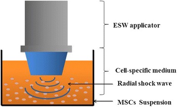
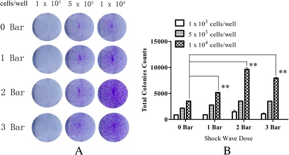
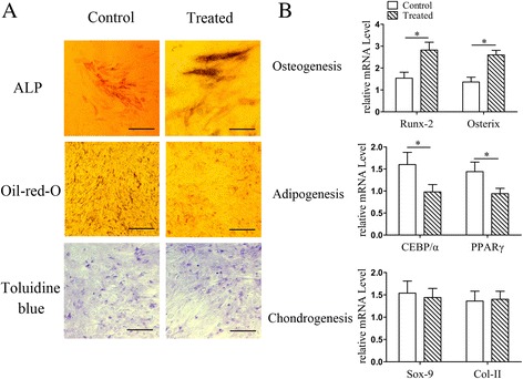
Methods: MSCs were cultured in vitro, harvested, resuspended, and treated with various doses of radial shockwaves in a floating system. Cell proliferation was evaluated by growth kinetics and Cell Counting Kit-8 (CCK-8) assay. In addition, the cell cycle and apoptotic activity were analyzed by fluorescence activated cell sorting. To explore the "stemness" of MSCs, cell colony-forming tests and multidifferentiation assays were performed. We also examined the MSC subcellular structure using transmission electron microscopy and examined the healing effects of these cells on cartilage defects by pathological analyses.

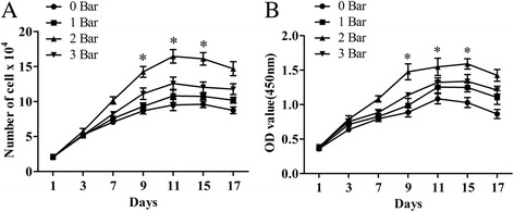
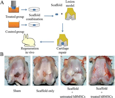
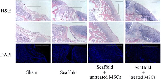
Results: The results of growth kinetics and CCK-8 assays showed that radial shockwave treatment significantly promoted MSC proliferation. Enhanced cell growth was also reflected by an increase in the numbers of cells in the S phase and a decrease in the numbers of cells arrested in the G0/G1 phase in shockwave-treated MSCs. Unexpectedly, shockwaves caused a slight increase in MSC apoptosis rates. Furthermore, radial shockwaves promoted self-replicating activity of MSCs. Transmission electron microscopy revealed that MSCs were metabolically activated by shockwave treatment. In addition, radial shockwaves favored MSC osteogenic differentiation but inhibited adipogenic activity. Most importantly, MSCs pretreated by radial shockwaves exhibited an enhanced healing effect on cartilage defects in vivo. Compared with control groups, shockwave-treated MSCs combined with bio-scaffolds significantly improved histological scores of injured rabbit knees.

Conclusions: In the present study, we found that radial shockwaves significantly promoted the proliferation and self-renewal of MSCs in vitro and safely accelerated the cartilage repair process in vivo, indicating favorable clinical outcomes.

Keywords: Cartilage repair; Mesenchymal stem cell; Radial shockwave.


Conflict of interest statement

Ethics approval

Informed consent was obtained from all patients for research purposes, and experiments were approved by the Ethics Review Committee of the PLA General Hospital. All animal experimental protocols were in compliance with the Animal Welfare Act and were approved by the Animal Care and Use Committee of the Laboratory Animal Research Center at the PLA General Hospital (Reference number: 2015-X11-10).

Consent for publication

Not applicable.

Competing interests

The authors declare that they have no competing interests.

Publisher’s Note

Springer Nature remains neutral with regard to jurisdictional claims in published maps and institutional affiliations.

Figures

Fig. 1
Fig. 1
Floating system of radial shockwave stimulation. MSCs harvested and resuspended in 25 ml of culture medium in 100-mm cell culture dishes received the energy of shockwaves directly. ESW extracorporeal shockwave, MSC mesenchymal stem cell
Fig. 2
Fig. 2
Radial shockwave stimulation promotes proliferation of MSCs in a dose-dependent manner. Trypan blue exclusion cell counting (a) and CCK-8-based cell proliferation assay (b) showed shockwave-treated MSCs exert stronger proliferative effects than control cells, and maximum cell viability occurred on the 11th day after stimulation. Optimal stimulation dose was 2 bars. Shockwave-treated MSCs incubated and measured at different pressures and compared with control groups in at least four independent experiments (different groups marked with different symbols). *Statistically significant difference compared with control group, P < 0.05
Fig. 3
Fig. 3
Radial shockwave influences cell cycle and apoptosis of MSC. Promotion effect reflected by cell cycle assays. Gray zones represent percentage of cells in the S phase. Higher percentage of shockwave-treated MSCs in the S phase compared to the control. On the basis of percentage of cells in the S phase and apoptosis, 2 bars is the optimal stimulation dose. Cell cycles detected by flow cytometry, and different pressures of shockwave-treated MSCs compared with control groups in at least four independent experiments. Representative data from a single experiment shown
Fig. 4
Fig. 4
Radial shockwaves promote colony-forming unit fibroblast formation of MSCs. a Comparisons of colony-formation efficiency. b Transformed data prepared using ImageJ software. Cell number in each subgroup shown above. When cell number is 1 × 10³ cells, there was no significant difference among the groups. There was a slight difference when cell number is 5 × 10³ cells. However, there is a significant difference when cell number is 1 × 10⁴ cells; the 2 bars group showed the highest colony-formation efficiency. CFU-F formation assay performed at least three times independently; representative data from a single experiment shown. **Statistically significant difference compared with control groups, P < 0.001
Fig. 5
Fig. 5
Radial shockwave treatment influence subcellular structure of MSCs. a Number of organelles increased. b In the treated group, Golgi apparatus and endoplasmic reticulum increased, and endoplasmic reticulum became increasingly well ordered and extended. c Nuclear diameter and area increased significantly, and kernel exhibited apparent chromosome replication. d Obvious increase in number of mitochondria in the treated group compared with the control. Scale bar: a 500 nm; b, d 200 nm; c 300 nm
Fig. 6
Fig. 6
Impact of radial shockwaves on multidifferentiation of MSCs. a Multidifferentiation tests of MSCs showed radial shockwave stimulation increased ALP activity but decreased formation of Oil Red-O-positive lipid droplets, indicating that shockwaves promote osteogenesis induction and block adipogenesis at the same time. No significant differences in the two cohorts of cells after chondrogenic induction. b Impact of shockwaves on MSC differentiation also reflected by mRNA levels of Runx-2, Osterix, CEBP/α, PPARγ, Sox-9, and Col-II. Quantitative PCR assay performed at least three times independently; representative result shown. *Statistically significant difference compared with control groups, P < 0.05. ALP alkaline phosphatase, Col-II collagen type II
Fig. 7
Fig. 7
Cartilage repair model and evaluation of gross repairing effect. a Flow diagram of cartilage repairing experiments. b Gross appearance in each group. Scale bar = 5 mm. hBMSC human bone marrow mesenchymal stem cell
Fig. 8
Fig. 8
Pathological analyses of radial-shockwave-treated MSCs mediated cartilage regeneration. H&E-stained section showing integrity of repaired tissue. Indicated areas magnified to show details. Using DAPI stain, number and morphology of nucleus shown. Cartilage cells markedly increased in shockwave-treated group compared to control group. Scale bar = 1 mm. H&E hematoxylin and eosin, DAPI 4′,6-diamidino-2-phenylindole, MSC mesenchymal stem cell
Fig. 9
Fig. 9
Radial shockwaves enhance formation of cartilage matrix and type II collagen in vivo. At 8 weeks following surgery, histochemical staining (Alcian blue and Safranin O/Fast Green) and immunohistochemical staining (type II collagen and PCNA) were performed. Stained sections scored using the modified ICRS II histology scoring system for cartilage repair. Scale bar = 1 mm. PCNA proliferating cell nuclear antigen, MSC mesenchymal stem cell

Similar articles

Cited by

References

    1. Bhattacharjee M, Coburn J, Centola M, Murab S, Barbero A, Kaplan DL, Martin I, Ghosh S. Tissue engineering strategies to study cartilage development, degeneration and regeneration. Adv Drug Deliv Rev. 2015;84:107–22. 10.1016/j.addr.2014.08.010. - PubMed
    1. de Windt TS, Hendriks JA, Zhao X, Vonk LA, Creemers LB, Dhert WJ, Randolph MA, Saris DB. Concise review: Unraveling stem cell cocultures in regenerative medicine: which cell interactions steer cartilage regeneration and how? Stem Cells Transl Med. 2014;3:723–733. doi: 10.5966/sctm.2013-0207. - DOI - PMC - PubMed
    1. Decker RS. Articular cartilage and joint development from embryogenesis to adulthood. Semin Cell Dev Biol. 2017;62:50–6. 10.1016/j.semcdb.2016.10.005. - PMC - PubMed
    1. Hui JH, Goyal D, Nakamura N, Ochi M, Asian Cartilage S. Cartilage repair. 2013 Asian update. Arthroscopy. 2013;29:1992–2000. doi: 10.1016/j.arthro.2013.06.009. - DOI - PubMed
    1. Anderson JA, Little D, Toth AP, Moorman CT, 3rd, Tucker BS, Ciccotti MG, Guilak F. Stem cell therapies for knee cartilage repair: the current status of preclinical and clinical studies. Am J Sports Med. 2014;42:2253–2261. doi: 10.1177/0363546513508744. - DOI - PMC - PubMed

Publication types