Clinical and laboratory predictors of Lassa fever outcome in a dedicated treatment facility in Nigeria: a retrospective, observational cohort study
- PMID: 29523497
- PMCID: PMC5984133
- DOI: 10.1016/S1473-3099(18)30121-X
Clinical and laboratory predictors of Lassa fever outcome in a dedicated treatment facility in Nigeria: a retrospective, observational cohort study
Erratum in
-
Corrections.Lancet Infect Dis. 2018 May;18(5):492. doi: 10.1016/S1473-3099(18)30208-1. Epub 2018 Mar 16. Lancet Infect Dis. 2018. PMID: 29555583 No abstract available.
Abstract
Background: Lassa fever is a viral haemorrhagic disease endemic to west Africa. No large-scale studies exist from Nigeria, where the Lassa virus (LASV) is most diverse. LASV diversity, coupled with host genetic and environmental factors, might cause differences in disease pathophysiology. Small-scale studies in Nigeria suggest that acute kidney injury is an important clinical feature and might be a determinant of survival. We aimed to establish the demographic, clinical, and laboratory factors associated with mortality in Nigerian patients with Lassa fever, and hypothesised that LASV was the direct cause of intrinsic renal damage for a subset of the patients with Lassa fever.
Methods: We did a retrospective, observational cohort study of consecutive patients in Nigeria with Lassa fever, who tested positive for LASV with RT-PCR, and were treated in Irrua Specialist Teaching Hospital. We did univariate and multivariate statistical analyses, including logistic regression, of all demographic, clinical, and laboratory variables available at presentation to identify the factors associated with patient mortality.
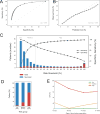
Findings: Of 291 patients treated in Irrua Specialist Teaching Hospital between Jan 3, 2011, and Dec 11, 2015, 284 (98%) had known outcomes (died or survived) and seven (2%) were discharged against medical advice. Overall case-fatality rate was 24% (68 of 284 patients), with a 1·4 times increase in mortality risk for each 10 years of age (p=0·00017), reaching 39% (22 of 57) for patients older than 50 years. Of 284 patients, 81 (28%) had acute kidney injury and 104 (37%) had CNS manifestations and thus both were considered important complications of acute Lassa fever in Nigeria. Acute kidney injury was strongly associated with poor outcome (case-fatality rate of 60% [49 of 81 patients]; odds ratio [OR] 15, p<0·00001). Compared with patients without acute kidney injury, those with acute kidney injury had higher incidence of proteinuria (32 [82%] of 39 patients) and haematuria (29 [76%] of 38) and higher mean serum potassium (4·63 [SD 1·04] mmol/L) and lower blood urea nitrogen to creatinine ratio (8·6 for patients without clinical history of fluid loss), suggesting intrinsic renal damage. Normalisation of creatinine concentration was associated with recovery. Elevated serum creatinine (OR 1·3; p=0·046), aspartate aminotransferase (OR 1·5; p=0·075), and potassium (OR 3·6; p=0·0024) were independent predictors of death.
Interpretation: Our study presents detailed clinical and laboratory data for Nigerian patients with Lassa fever and provides strong evidence for intrinsic renal dysfunction in acute Lassa fever. Early recognition and treatment of acute kidney injury might significantly reduce mortality.
Funding: German Research Foundation, German Center for Infection Research, Howard Hughes Medical Institute, US National Institutes of Health, and World Bank.
Copyright © 2018 Elsevier Ltd. All rights reserved.
Conflict of interest statement
All authors declare not having any conflicts of interest.
Figures


Comment in
-
Predicting outcome and improving treatment for Lassa fever.Lancet Infect Dis. 2018 Jun;18(6):594-595. doi: 10.1016/S1473-3099(18)30116-6. Epub 2018 Mar 6. Lancet Infect Dis. 2018. PMID: 29523495 No abstract available.
-
Early detection of Lassa fever: the need for point-of-care diagnostics.Lancet Infect Dis. 2018 Jun;18(6):601-602. doi: 10.1016/S1473-3099(18)30277-9. Lancet Infect Dis. 2018. PMID: 29856352 No abstract available.
References
-
- Grant DS, Khan H, Schieffelin J, Bausch DG. Chapter 4 - Lassa Fever Emerging Infectious Diseases. Amsterdam: Academic Press; 2014. pp. 37–59.
-
- Frame JD, Baldwin JM, Jr, Gocke DJ, Troup JM. Lassa fever, a new virus disease of man from West Africa. I. Clinical description and pathological findings. Am J Trop Med Hyg. 1970;19(4):670–6. - PubMed
-
- McCormick JB, King IJ, Webb PA, et al. Lassa Fever: Effective Therapy with Ribavirin. New England Journal of Medicine. 1986;314(1):20–6. - PubMed
Publication types
MeSH terms
Grants and funding
LinkOut - more resources
Full Text Sources
Other Literature Sources
Research Materials

