Comparative Study
. 2018 May 1;84(10):e00029-18.
doi: 10.1128/AEM.00029-18. Print 2018 May 15.

Tissue Localization and Variation of Major Symbionts in Haemaphysalis longicornis, Rhipicephalus haemaphysaloides, and Dermacentor silvarum in China

Affiliations
Comparative Study

Tissue Localization and Variation of Major Symbionts in Haemaphysalis longicornis, Rhipicephalus haemaphysaloides, and Dermacentor silvarum in China

Mengfei Wang et al. Appl Environ Microbiol. .

Abstract

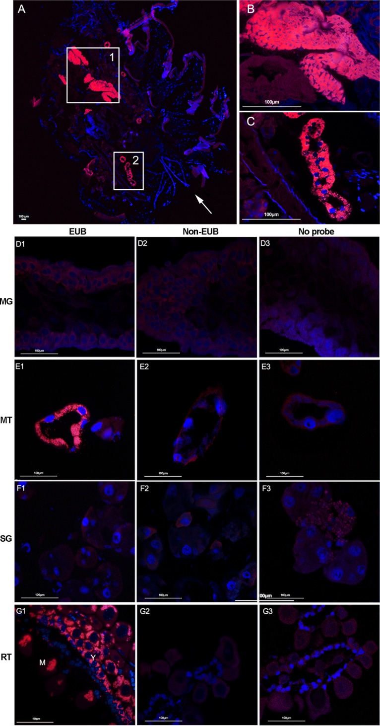
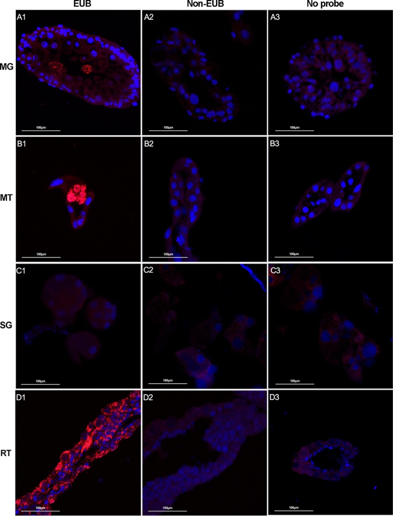
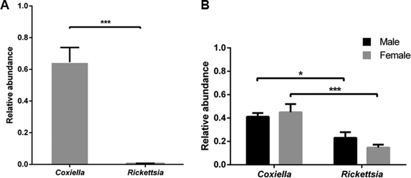
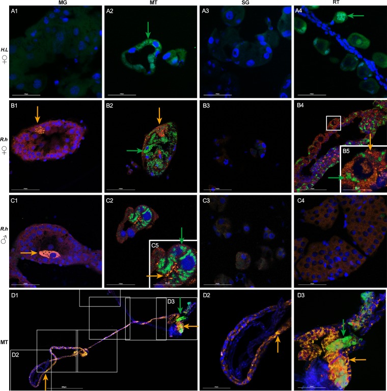
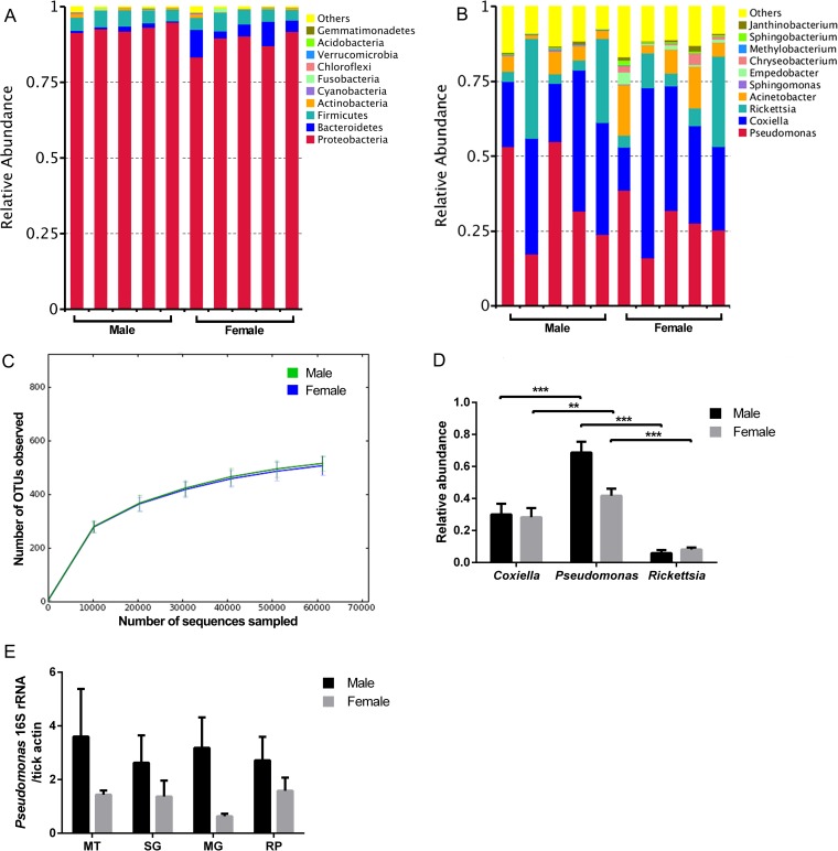
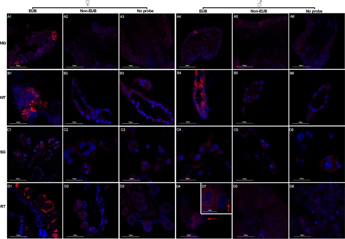
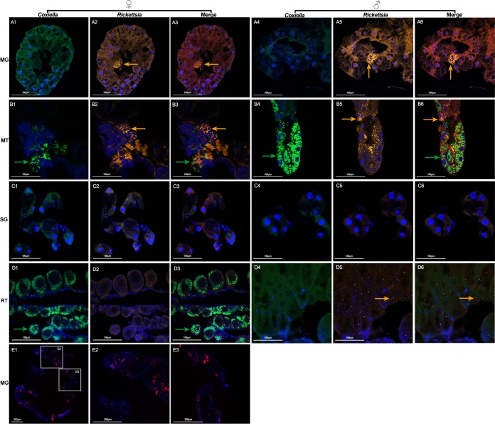
Ticks are important disease vectors, as they transmit a variety of human and animal pathogens worldwide. Symbionts that coevolved with ticks confer crucial benefits to their host in nutrition metabolism, fecundity, and vector competence. Although over 100 tick species have been identified in China, general information on tick symbiosis is limited. Here, we visualized the tissue distribution of Coxiella sp. and Rickettsia sp. in lab-reared Haemaphysalis longicornis and Rhipicephalus haemaphysaloides by fluorescent in situ hybridization. We found that Coxiella sp. colonized exclusively the Malpighian tubules and ovaries of H. longicornis, while Rickettsia sp. additionally colonized the midgut of R. haemaphysaloides We also investigated the population structure of microbiota in Dermacentor silvarum ticks collected from Inner Mongolia, China, and found that Coxiella, Rickettsia, and Pseudomonas are the three dominant genera. No significant difference in microbiota composition was found between male and female D. silvarum ticks. We again analyzed the tissue localization of Coxiella sp. and Rickettsia sp. and found that they displayed tissue tropisms similar to those in R. haemaphysaloides, except that Rickettsia sp. colonized the nuclei of spermatids instead of ovaries in D. silvarum Altogether, our results suggest that Coxiella sp. and Rickettsia sp. are the main symbionts in the three ticks and reside primarily in midgut, Malpighian tubules, and reproductive tissues, but their tissue distribution varies in association with species and sexes.IMPORTANCE Tick-borne diseases constitute a major public health burden, as they are increasing in frequency and severity worldwide. The presence of symbionts helps ticks to metabolize nutrients, promotes fecundity, and influences pathogen infections. Increasing numbers of tick-borne pathogens have been identified in China; however, knowledge of native ticks, especially tick symbiosis, is limited. In this study, we analyze the distribution of Coxiella sp. and Rickettsia sp. in tissues of laboratory-reared Haemaphysalis longicornis and Rhipicephalus haemaphysaloides and field-collected Dermacentor silvarum We found that the localization patterns of Coxiella sp. in three Chinese tick species were similar to those of other tick species. We also found a previously undefined intracellular localization of Rickettsia sp. in tick midgut and spermatids. In addition, we demonstrate that tissue tropisms of symbionts vary between species and sexes. Our findings provide new insights into the tissue localization of symbionts in native Chinese ticks and pave the way for further understanding of their functional capabilities and symbiotic interactions with ticks.

Keywords: Coxiella; Rickettsia; microbiota; ticks; tissue localization.


Figures

FIG 1
FIG 1
Localization of microbiota in whole body (A to C) and different tissues (D to G) of female H. longicornis by FISH analysis with universal 16S rRNA probe (red). Nuclei were stained with DAPI (blue). (A) Signals detected in whole body sections; the arrow denotes the direction from head to bottom; (B) close-up view of box 1 showing the ovary filled with microbiota; (C) close-up view of box 2 showing that the microbiota colonized the Malpighian tubules. Images are representative of at least 5 individual tick sections. Different tissues, including midgut (D), Malpighian tubules (E), salivary gland (F), and ovaries (G) were hybridized with universal 16S rRNA probe (EUB) (D1, E1, F1, and G1), noneubacterial probe (Non-EUB) (D2, E2, F2, and G2), and no probe (D3, E3, F3, and G3). MG, midgut; MT, Malpighian tubules; SG, salivary glands; RT, ovaries; Y, young oocytes; M, mature oocytes. Tissues from at least 5 individual ticks were pooled and used for FISH analysis. Images are representative of three independent experiments.
FIG 2
FIG 2
Localization of microbiota in different tissues of female R. haemaphysaloides by FISH analysis with universal 16S rRNA probe (red). Nuclei were stained with DAPI (blue). Fluorescent signals were examined in midgut (A), Malpighian tubules (B), salivary glands (C), and ovaries (D). Hybridizations with noneubacterial probe (Non-EUB) and without probe were used as negative controls. Tissues from at least 5 individual ticks were pooled and used for FISH analysis. Images are representative of three independent experiments.
FIG 3
FIG 3
Relative abundance of Coxiella sp. and Rickettsia sp. in H. longicornis (A) and R. haemaphysaloides (B) determined by real-time quantitative PCR. Error bars indicate standard errors (n = 15). Significance was determined by Mann-Whitney test. *, P < 0.05; ***, P < 0.001.
FIG 4
FIG 4
Localization of Coxiella sp. (green) and Rickettsia sp. (yellow) in H. longicornis (A) and R. haemaphysaloides (B to D) using species specific probes. Nuclei were stained with DAPI (blue). Merged images of DAPI and Coxiella sp. staining in female H. longicornis (A1 to A4). Merged images of DAPI, Coxiella sp., and Rickettsia sp. staining in female (B1 to B4) and male (C1 to C4) R. haemaphysaloides. Spliced view of whole-mount in situ hybridization in female Malpighian tubules of R. haemaphysaloides (D1 to D3). (A1, B1, C1), midgut (MG); (A2, B2, C2), Malpighian tubules (MT); (A3, B3, C3), salivary glands (SG); (A4), ovaries of H. longicornis (reproductive tissues [RT]); (B4), ovaries of R. haemaphysaloides; (C4), testes of R. haemaphysaloides; (B5) close-up view of boxed region, showing colocalization of Rickettsia sp. and Coxiella sp. in ovaries; (C5) close-up view of panel C2, colocalization of Rickettsia sp. and Coxiella sp. in Malpighian tubules. (D2, D3) Close-up view of boxed regions in D1. Green arrows denote Coxiella sp. and yellow arrows denote Rickettsia sp. Tissues from at least 5 individual ticks were pooled and used for FISH analysis. Each image shows a single focal plane. Images are representative of three independent experiments. Bars, 100 μm.
FIG 5
FIG 5
Population community of D. silvarum by 16S rRNA pyrosequencing. Phylum level (A) and genus level (B) abundance profiles for individual D. silvarum males and females prior to a blood meal. Each column represents one tick. (C) Rarefaction curve of the 16S rRNA gene sequences in male and female D. silvarum ticks based on OTUs determined at 97% similarity. (D) Relative abundance of Coxiella sp., Pseudomonas sp., and Rickettsia sp. in D. silvarum analyzed by qPCR. (E) Quantification of Pseudomonas sp. in different tick tissues by qPCR. Tissues from five individual tick were pooled for one biological replicate. Five biological replicates were used for qPCR analysis. Error bars indicate standard errors (n = 5). Statistical significance was determined using the Mann-Whitney test. **, P < 0.01; ***, P < 0.001.
FIG 6
FIG 6
Localization of microbiota in different tissues of female and male D. silvarum ticks by FISH analysis with universal 16S rRNA probe (red). Nuclei were stained with DAPI (blue). Fluorescent signals were examined in midgut (A), Malpighian tubules (B), salivary glands (C), ovaries (D1 to D3), and testes (D4 to D7). Hybridizations with noneubacterial probe (Non-EUB) and without probe were used as negative controls. Red arrows denote residential bacteria. MG, midgut; MT, Malpighian tubules; SG, salivary glands; RT, reproductive tissue. Tissues from at least 5 individual ticks were pooled and used for FISH analysis. Images are representative of three independent experiments.
FIG 7
FIG 7
Costaining of Coxiella sp. (green), Rickettsia sp. (yellow), and Pseudomonas sp. (red) in D. silvarum. Nuclei were stained with DAPI (blue). Sections of midgut (A1 to A6), Malpighian tubules (B1 to B6), salivary glands (C1 to C6), ovaries (D1 to D3), and testes (D4 to D6) were hybridized with Coxiella sp.-specific 23S rRNA probe (A1 and A4, B1 and B4, C1 and C4, and D1 and D4) and Rickettsia sp.-specific 16S rRNA probe (A2 and A5, B2 and B5, C2 and C5, and D2 and D5) simultaneously. (A3 and A6, B3 and B6, C3 and C6, and D3 and D6) Merged images of DAPI, Coxiella sp., and Rickettsia sp. staining. Green arrows denote Coxiella sp. and yellow arrows denote Rickettsia sp. MG, midgut; MT, Malpighian tubules; SG, salivary glands; RT, reproductive tissue. (E1 to E3) Whole-mount in situ hybridization of D. silvarum midgut using Pseudomonas sp.-specific 16S rRNA probe. (E2, E3) Close-up view of boxed regions in panel E1. Tissues from at least 5 individual ticks were pooled and used for FISH analysis. Each image shows a single focal plane. Images are representative of three independent experiments.

References

    1. WHO. 2017. Global vector control response 2017–2030. World Health Organization, Geneva, Switzerland.
    1. Fang LQ, Liu K, Li XL, Liang S, Yang Y, Yao HW, Sun RX, Sun Y, Chen WJ, Zuo SQ, Ma MJ, Li H, Jiang JF, Liu W, Yang XF, Gray GC, Krause PJ, Cao WC. 2015. Emerging tick-borne infections in mainland China: an increasing public health threat. Lancet Infect Dis 15:1467–1479. doi: 10.1016/S1473-3099(15)00177-2. - DOI - PMC - PubMed
    1. Wu XB, Na RH, Wei SS, Zhu JS, Peng HJ. 2013. Distribution of tick-borne diseases in China. Parasit Vectors 6:119. doi: 10.1186/1756-3305-6-119. - DOI - PMC - PubMed
    1. Zhou X, Xia S, Huang JL, Tambo E, Zhuge HX, Zhou XN. 2014. Human babesiosis, an emerging tick-borne disease in the People's Republic of China. Parasit Vectors 7:509. doi: 10.1186/s13071-014-0509-3. - DOI - PMC - PubMed
    1. Chen Z, Yang X, Bu F, Yang X, Yang X, Liu J. 2010. Ticks (Acari: Ixodoidea: Argasidae, Ixodidae) of China. Exp Appl Acarol 51:393–404. doi: 10.1007/s10493-010-9335-2. - DOI - PubMed

Publication types

MeSH terms