

On the nature and function of organizers
- PMID: 29523654
- PMCID: PMC5868996
- DOI: 10.1242/dev.159525
On the nature and function of organizers
Abstract
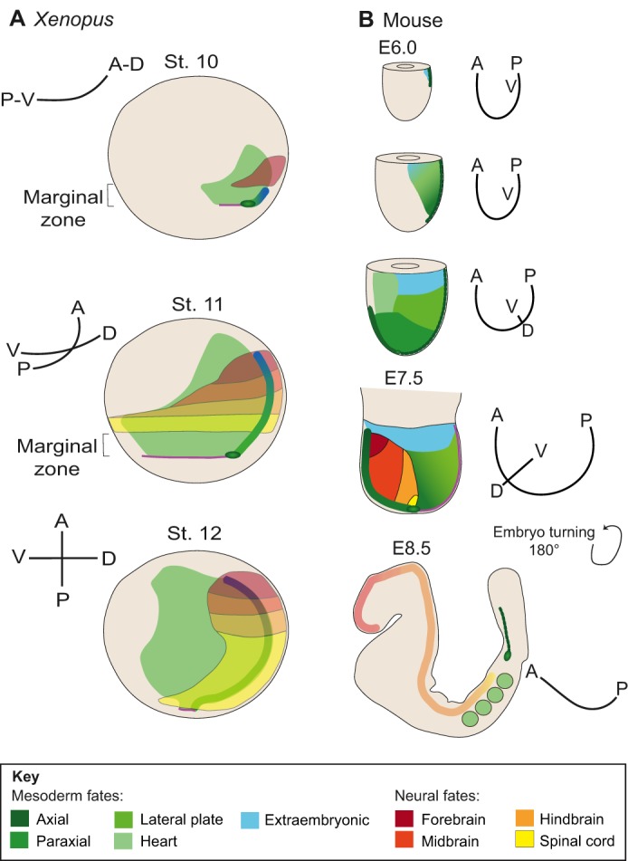
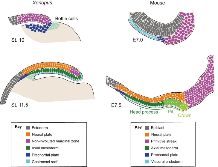
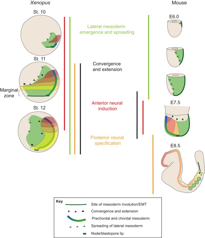
Organizers, which comprise groups of cells with the ability to instruct adjacent cells into specific states, represent a key principle in developmental biology. The concept was first introduced by Spemann and Mangold, who showed that there is a cellular population in the newt embryo that elicits the development of a secondary axis from adjacent cells. Similar experiments in chicken and rabbit embryos subsequently revealed groups of cells with similar instructive potential. In birds and mammals, organizer activity is often associated with a structure known as the node, which has thus been considered a functional homologue of Spemann's organizer. Here, we take an in-depth look at the structure and function of organizers across species and note that, whereas the amphibian organizer is a contingent collection of elements, each performing a specific function, the elements of organizers in other species are dispersed in time and space. This observation urges us to reconsider the universality and meaning of the organizer concept.
Keywords: Axial organization; Body plan; Neural induction; Organizer; Spemann; Vertebrate embryo.
© 2018. Published by The Company of Biologists Ltd.
Conflict of interest statement
Competing interestsThe authors declare no competing or financial interests.
Figures
Similar articles
-
Spemann's organizer and self-regulation in amphibian embryos.Nat Rev Mol Cell Biol. 2006 Apr;7(4):296-302. doi: 10.1038/nrm1855. Nat Rev Mol Cell Biol. 2006. PMID: 16482093 Free PMC article. Review.
-
Introducing the Spemann-Mangold organizer: experiments and insights that generated a key concept in developmental biology.Int J Dev Biol. 2001;45(1):1-11. Int J Dev Biol. 2001. PMID: 11291840
-
Microarray analysis of Foxa2 mutant mouse embryos reveals novel gene expression and inductive roles for the gastrula organizer and its derivatives.BMC Genomics. 2008 Oct 30;9:511. doi: 10.1186/1471-2164-9-511. BMC Genomics. 2008. PMID: 18973680 Free PMC article.
-
Head-organizing activities of endodermal tissues in vertebrates.Cell Mol Biol (Noisy-le-grand). 1999 Jul;45(5):481-92. Cell Mol Biol (Noisy-le-grand). 1999. PMID: 10512181 Review.
-
The avian organizer.Int J Dev Biol. 2001;45(1):281-7. Int J Dev Biol. 2001. PMID: 11291858 Review.
Cited by
-
The Organizer and Its Signaling in Embryonic Development.J Dev Biol. 2021 Nov 1;9(4):47. doi: 10.3390/jdb9040047. J Dev Biol. 2021. PMID: 34842722 Free PMC article. Review.
-
Neural induction drives body axis formation during embryogenesis, but a neural induction-like process drives tumorigenesis in postnatal animals.Front Cell Dev Biol. 2023 May 9;11:1092667. doi: 10.3389/fcell.2023.1092667. eCollection 2023. Front Cell Dev Biol. 2023. PMID: 37228646 Free PMC article. Review.
-
Development of node architecture and emergence of molecular organizer characteristics in the pig embryo.Dev Dyn. 2025 Aug;254(8):916-934. doi: 10.1002/dvdy.715. Epub 2024 May 11. Dev Dyn. 2025. PMID: 38733144 Free PMC article.
-
Cell lineage-guided mass spectrometry reveals increased energy metabolism and reactive oxygen species in the vertebrate organizer.Proc Natl Acad Sci U S A. 2024 Feb 6;121(6):e2311625121. doi: 10.1073/pnas.2311625121. Epub 2024 Feb 1. Proc Natl Acad Sci U S A. 2024. PMID: 38300871 Free PMC article.
-
Minimal Developmental Computation: A Causal Network Approach to Understand Morphogenetic Pattern Formation.Entropy (Basel). 2022 Jan 10;24(1):107. doi: 10.3390/e24010107. Entropy (Basel). 2022. PMID: 35052133 Free PMC article.
References
-
- Bachvarova R. F., Skromne I. and Stern C. D. (1998). Induction of primitive streak and Hensen's node by the posterior marginal zone in the early chick embryo. Development 125, 3521-3534. - PubMed
Publication types
MeSH terms
Grants and funding
LinkOut - more resources
Full Text Sources
Other Literature Sources
