

Does the microbiome and virome contribute to myalgic encephalomyelitis/chronic fatigue syndrome?
- PMID: 29523751
- PMCID: PMC5843715
- DOI: 10.1042/CS20171330
Does the microbiome and virome contribute to myalgic encephalomyelitis/chronic fatigue syndrome?
Abstract
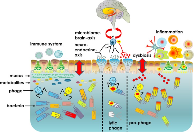
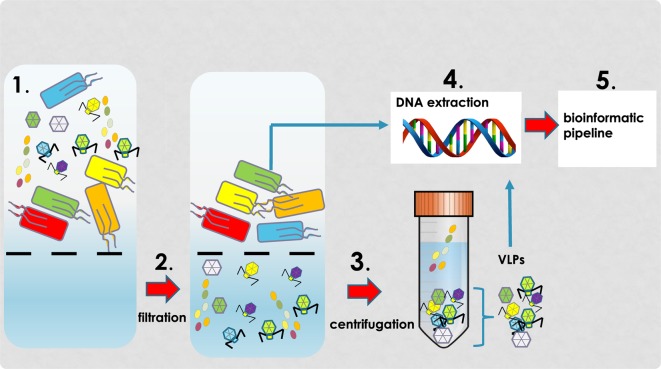
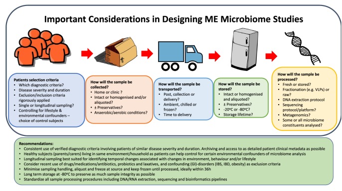
Myalgic encephalomyelitis (ME)/chronic fatigue syndrome (CFS) (ME/CFS) is a disabling and debilitating disease of unknown aetiology. It is a heterogeneous disease characterized by various inflammatory, immune, viral, neurological and endocrine symptoms. Several microbiome studies have described alterations in the bacterial component of the microbiome (dysbiosis) consistent with a possible role in disease development. However, in focusing on the bacterial components of the microbiome, these studies have neglected the viral constituent known as the virome. Viruses, particularly those infecting bacteria (bacteriophages), have the potential to alter the function and structure of the microbiome via gene transfer and host lysis. Viral-induced microbiome changes can directly and indirectly influence host health and disease. The contribution of viruses towards disease pathogenesis is therefore an important area for research in ME/CFS. Recent advancements in sequencing technology and bioinformatics now allow more comprehensive and inclusive investigations of human microbiomes. However, as the number of microbiome studies increases, the need for greater consistency in study design and analysis also increases. Comparisons between different ME/CFS microbiome studies are difficult because of differences in patient selection and diagnosis criteria, sample processing, genome sequencing and downstream bioinformatics analysis. It is therefore important that microbiome studies adopt robust, reproducible and consistent study design to enable more reliable and valid comparisons and conclusions to be made between studies. This article provides a comprehensive review of the current evidence supporting microbiome alterations in ME/CFS patients. Additionally, the pitfalls and challenges associated with microbiome studies are discussed.
Keywords: Myalgic Encephalomyelitis/Chronic Fatigue Syndrome; Virome; microbiome.
© 2018 The Author(s).
Conflict of interest statement
The authors declare that there are no competing interests associated with the manuscript.
Figures
Similar articles
-
The potential therapeutic approaches targeting gut health in myalgic encephalomyelitis/chronic fatigue syndrome (ME/CFS): a narrative review.J Transl Med. 2025 May 11;23(1):530. doi: 10.1186/s12967-025-06527-x. J Transl Med. 2025. PMID: 40350437 Free PMC article. Review.
-
The Gut Microbiome in Myalgic Encephalomyelitis (ME)/Chronic Fatigue Syndrome (CFS).Front Immunol. 2022 Jan 3;12:628741. doi: 10.3389/fimmu.2021.628741. eCollection 2021. Front Immunol. 2022. PMID: 35046929 Free PMC article. Review.
-
A systematic review of enteric dysbiosis in chronic fatigue syndrome/myalgic encephalomyelitis.Syst Rev. 2018 Dec 20;7(1):241. doi: 10.1186/s13643-018-0909-0. Syst Rev. 2018. PMID: 30572962 Free PMC article.
-
Reduced diversity and altered composition of the gut microbiome in individuals with myalgic encephalomyelitis/chronic fatigue syndrome.Microbiome. 2016 Jun 23;4(1):30. doi: 10.1186/s40168-016-0171-4. Microbiome. 2016. PMID: 27338587 Free PMC article.
-
Changes in Gut and Plasma Microbiome following Exercise Challenge in Myalgic Encephalomyelitis/Chronic Fatigue Syndrome (ME/CFS).PLoS One. 2015 Dec 18;10(12):e0145453. doi: 10.1371/journal.pone.0145453. eCollection 2015. PLoS One. 2015. PMID: 26683192 Free PMC article.
Cited by
-
Finding intestinal fortitude: Integrating the microbiome into a holistic view of depression mechanisms, treatment, and resilience.Neurobiol Dis. 2020 Feb;135:104578. doi: 10.1016/j.nbd.2019.104578. Epub 2019 Aug 24. Neurobiol Dis. 2020. PMID: 31454550 Free PMC article. Review.
-
Oral Bacteriotherapy Reduces the Occurrence of Chronic Fatigue in COVID-19 Patients.Front Nutr. 2022 Jan 12;8:756177. doi: 10.3389/fnut.2021.756177. eCollection 2021. Front Nutr. 2022. PMID: 35096923 Free PMC article.
-
Deep phenotyping of myalgic encephalomyelitis/chronic fatigue syndrome in Japanese population.Sci Rep. 2020 Nov 16;10(1):19933. doi: 10.1038/s41598-020-77105-y. Sci Rep. 2020. PMID: 33199820 Free PMC article.
-
The potential therapeutic approaches targeting gut health in myalgic encephalomyelitis/chronic fatigue syndrome (ME/CFS): a narrative review.J Transl Med. 2025 May 11;23(1):530. doi: 10.1186/s12967-025-06527-x. J Transl Med. 2025. PMID: 40350437 Free PMC article. Review.
-
The Role of Chronic Fatigue in Patients with Crohn's Disease.Life (Basel). 2023 Aug 5;13(8):1692. doi: 10.3390/life13081692. Life (Basel). 2023. PMID: 37629549 Free PMC article. Review.
References
Publication types
MeSH terms
Grants and funding
LinkOut - more resources
Full Text Sources
Other Literature Sources
Medical