

ELF3 promotes epithelial-mesenchymal transition by protecting ZEB1 from miR-141-3p-mediated silencing in hepatocellular carcinoma
- PMID: 29523781
- PMCID: PMC5845010
- DOI: 10.1038/s41419-018-0399-y
ELF3 promotes epithelial-mesenchymal transition by protecting ZEB1 from miR-141-3p-mediated silencing in hepatocellular carcinoma
Abstract
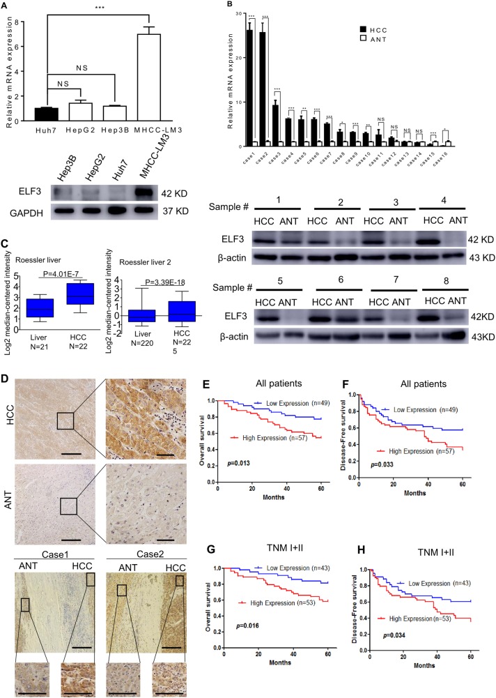
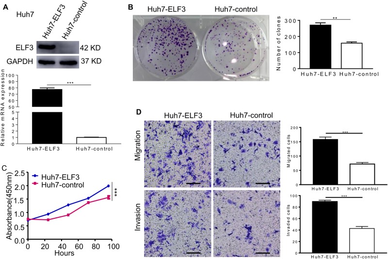
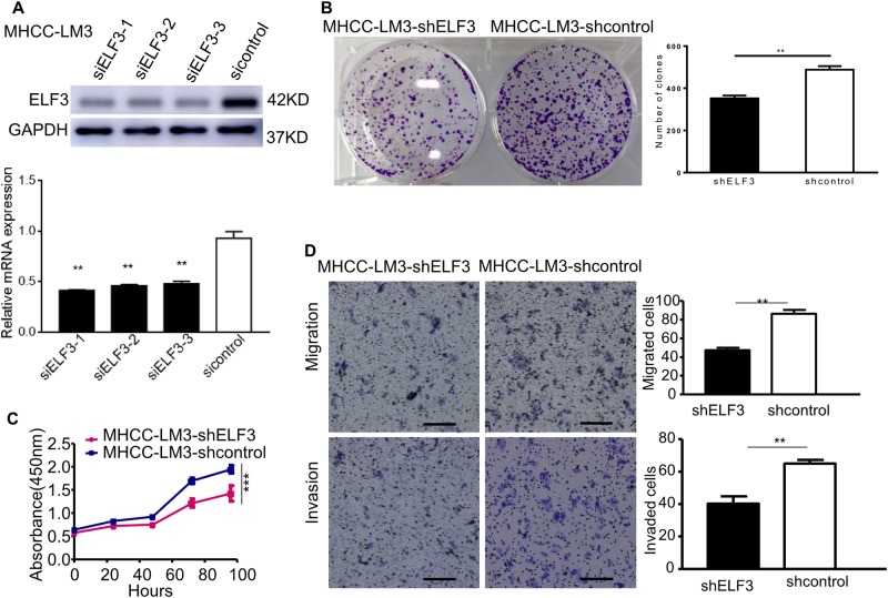
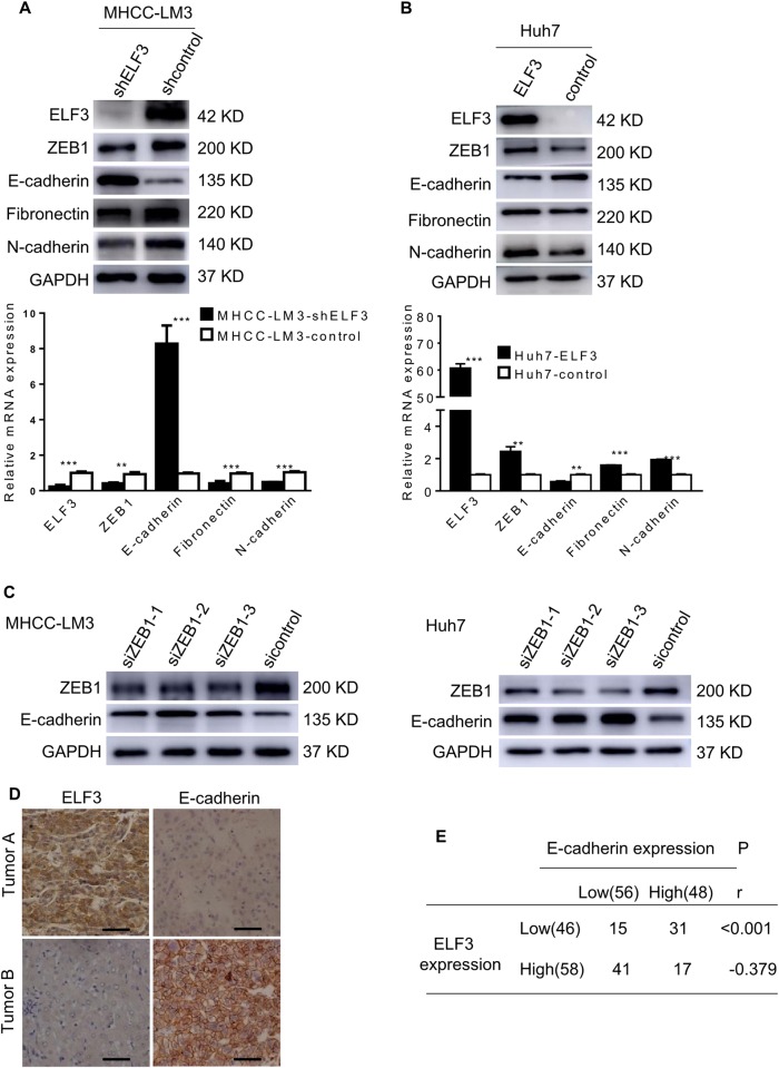
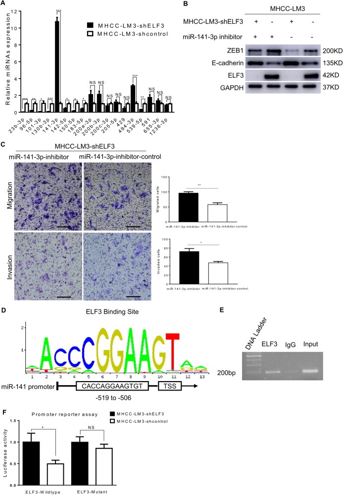
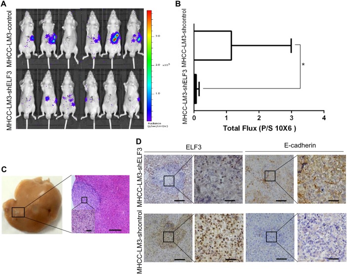
Hepatocellular carcinoma (HCC) is one of the most common malignant cancers and currently the third leading cause of cancer-related deaths, worldwide. Epithelial-mesenchymal transition (EMT) plays a major role in HCC progression. In this study, we first found that the expression of E74-like ETS transcription factor 3 (ELF3), a member of the E-twenty-six family of transcription factors, was increased in HCC tissues, and that ELF3 overexpression was associated with poor prognoses for HCC patients. Gain-of-function and loss-of-function studies revealed that increased ELF3 expression promoted HCC cell proliferation, migration, and invasion, while these processes were inhibited when ELF3 was silenced. Additionally, ELF3 was found to promote EMT, which we demonstrated through decreased E-cadherin expression and increased N-cadherin and fibronectin expression. ELF3 knockdown reversed EMT via repressing ZEB1 expression through miR-141-3p upregulation. Chromatin immunoprecipitation assays revealed that ELF3 bound to the miR-141-3p promoter, suppressing miR-141-3p expression. Taken together, our data show that ELF3 repressed E-cadherin and promoted EMT in HCC cells by suppressing miR-141-3p, thereby activating ZEB1. Thus, ELF3 may be a potential prognostic biomarker and/or therapeutic target for HCC.
Conflict of interest statement
The authors declare that they have no conflict of interest.
Figures
Similar articles
-
lncRNA TUG1-Mediated Mir-142-3p Downregulation Contributes to Metastasis and the Epithelial-to-Mesenchymal Transition of Hepatocellular Carcinoma by Targeting ZEB1.Cell Physiol Biochem. 2018;48(5):1928-1941. doi: 10.1159/000492517. Epub 2018 Aug 9. Cell Physiol Biochem. 2018. PMID: 30092578
-
miR-139-5p inhibits epithelial-mesenchymal transition, migration and invasion of hepatocellular carcinoma cells by targeting ZEB1 and ZEB2.Biochem Biophys Res Commun. 2015 Jul 31;463(3):315-21. doi: 10.1016/j.bbrc.2015.05.062. Epub 2015 May 27. Biochem Biophys Res Commun. 2015. PMID: 26022123
-
LncRNA HULC enhances epithelial-mesenchymal transition to promote tumorigenesis and metastasis of hepatocellular carcinoma via the miR-200a-3p/ZEB1 signaling pathway.Oncotarget. 2016 Jul 5;7(27):42431-42446. doi: 10.18632/oncotarget.9883. Oncotarget. 2016. PMID: 27285757 Free PMC article.
-
miR-200c: a versatile watchdog in cancer progression, EMT, and drug resistance.J Mol Med (Berl). 2016 Jun;94(6):629-44. doi: 10.1007/s00109-016-1420-5. Epub 2016 Apr 20. J Mol Med (Berl). 2016. PMID: 27094812 Review.
-
The function of the ELF3 gene and its mechanism in cancers.Life Sci. 2024 Jun 1;346:122637. doi: 10.1016/j.lfs.2024.122637. Epub 2024 Apr 12. Life Sci. 2024. PMID: 38614305 Review.
Cited by
-
Chemotherapy-induced miR-141/MAP4K4 signaling suppresses progression of colorectal cancer.Biosci Rep. 2018 Dec 21;38(6):BSR20180978. doi: 10.1042/BSR20180978. Print 2018 Dec 21. Biosci Rep. 2018. PMID: 30429233 Free PMC article.
-
Interpretation of Euphorbia Kansui Stir-Fried with Vinegar Treating Malignant Ascites by a UPLC-Q-TOF/MS Based Rat Serum and Urine Metabolomics Strategy Coupled with Network Pharmacology.Molecules. 2018 Dec 7;23(12):3246. doi: 10.3390/molecules23123246. Molecules. 2018. PMID: 30544627 Free PMC article.
-
miR-486-3p mediates hepatocellular carcinoma sorafenib resistance by targeting FGFR4 and EGFR.Cell Death Dis. 2020 Apr 20;11(4):250. doi: 10.1038/s41419-020-2413-4. Cell Death Dis. 2020. PMID: 32313144 Free PMC article.
-
The Mechanism of miR-141 Regulating the Proliferation and Metastasis of Liver Cancer Cells by Targeting STAT4.J Oncol. 2021 Oct 12;2021:5425491. doi: 10.1155/2021/5425491. eCollection 2021. J Oncol. 2021. PMID: 34675977 Free PMC article.
-
Metabolome Profiling of Malignant Ascites Identifies Candidate Metabolic Biomarkers of Hepatocellular Carcinoma.Curr Med Chem. 2024;31(13):1769-1780. doi: 10.2174/0929867330666230324153552. Curr Med Chem. 2024. PMID: 38666505
References
-
- Pan HW, et al. Overexpression of osteopontin is associated with intrahepatic metastasis, early recurrence, and poorer prognosis of surgically resected hepatocellular carcinoma. Cancer. 2003;98:119–127. - PubMed
-
- Polyak K, Weinberg RA. Transitions between epithelial and mesenchymal states: acquisition of malignant and stem cell traits. Nat. Rev. Cancer. 2009;9:265–273. - PubMed
-
- Hanahan D, Weinberg RA. The hallmarks of cancer. Cell. 2000;100:57–70. - PubMed
Publication types
MeSH terms
Substances
LinkOut - more resources
Full Text Sources
Other Literature Sources
Medical
Research Materials
