

PSMA expression: a potential ally for the pathologist in prostate cancer diagnosis
- PMID: 29523813
- PMCID: PMC5844862
- DOI: 10.1038/s41598-018-22594-1
PSMA expression: a potential ally for the pathologist in prostate cancer diagnosis
Abstract
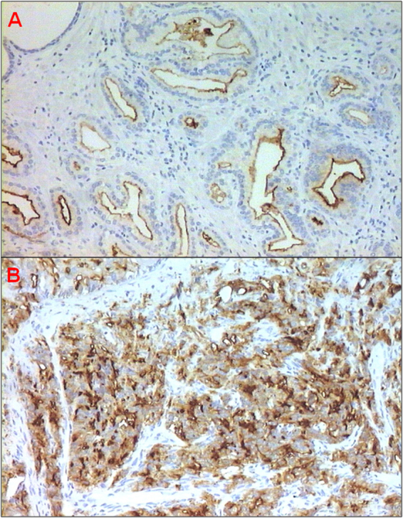
Prostate cancer (PCa) patients are risk-stratified on the basis of clinical stage and PSA level at diagnosis and the Gleason Score (GS) in prostate biopsy. However, these parameters are not completely accurate in discriminating between high- and low-risk disease, creating a need for a reliable marker to determine aggressiveness. Prostate-specific membrane antigen (PSMA) appears to fulfill this need. We analyzed 79 prostate biopsies and 28 prostatectomies to assess whether PSMA expression detected by immunohistochemistry is related to GS. PSMA expression was correlated with GS in both sample types (biopsies, P < 0.0001 and prostatectomy samples, P = 0.007). We observed lower PSMA expression in Gleason pattern 3 than Gleason pattern 4, suggesting that this biomarker could be useful to distinguish between these entities (p < 0.0001). The best cut-off value of 45% immunopositivity was determined by receiver operating characteristic (ROC) curve analysis. In Gleason pattern 3 vs. Gleason pattern 4 and 5, PSMA sensitivity was 84.1% (95% CI 76.5%-91.7%) and specificity was 95.2% (95% CI 90.6%-99.8%), with an area under the curve of 93.1 (95% CI 88.8-97.4). Our results suggest that PSMA represents a potential ally for the pathologist in the diagnostic work-up of PCa to overcome long-standing morphological classification limits.
Conflict of interest statement
The authors declare no competing interests.
Figures

References
-
- http://globocan.iarc.fr/old/FactSheets/cancers/prostate-new.asp, GLOBOCAN2012 (IARC), Section of Cancer Surveillance (20/2/2018).
-
- Epstein JI, et al. The 2014 International Society of Urological Pathology (ISUP) Consensus Conference on Gleason Grading of Prostatic Carcinoma: Definition of Grading Patterns and Proposal for a New Grading System. Am J Surg Pathol. 2016;40:244–52. - PubMed
-
- Israeli RS, Powell CT, Corr JG, Fair WR, Heston WD. Expression of the prostate-specific membrane antigen. Cancer Res. 1994;54:1807–1811. - PubMed
Publication types
MeSH terms
Substances
LinkOut - more resources
Full Text Sources
Other Literature Sources
Medical
Research Materials
Miscellaneous
