

Interferon-β deficiency at asthma exacerbation promotes MLKL mediated necroptosis
- PMID: 29523863
- PMCID: PMC5844912
- DOI: 10.1038/s41598-018-22557-6
Interferon-β deficiency at asthma exacerbation promotes MLKL mediated necroptosis
Abstract
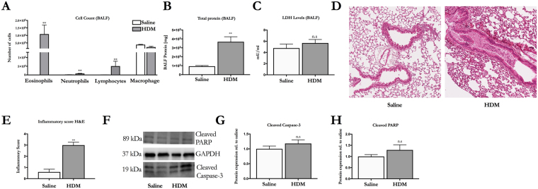
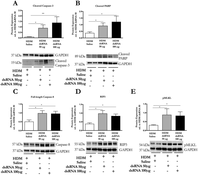
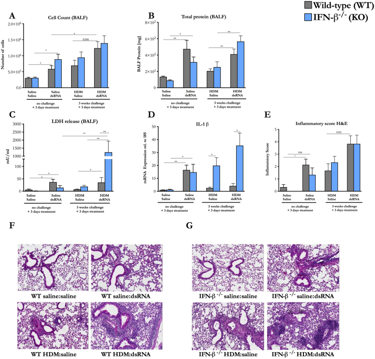
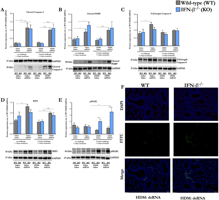
Defective production of antiviral interferon (IFN)-β is thought to contribute to rhinovirus-induced asthma exacerbations. These exacerbations are associated with elevated lung levels of lactate dehydrogenase (LDH), indicating occurrence of cell necrosis. We thus hypothesized that reduced lung IFN-β could contribute to necrotic cell death in a model of asthma exacerbations. Wild-type and IFN-β-/- mice were given saline or house dust mite (HDM) intranasally for 3 weeks to induce inflammation. Double-stranded RNA (dsRNA) was then given for additional 3 days to induce exacerbation. HDM induced an eosinophilic inflammation, which was not associated with increased expression of cleaved caspase-3, cleaved PARP or elevated bronchoalveolar lavage fluid (BALF) LDH levels in wild-type. However, exacerbation evoked by HDM + dsRNA challenges increased BALF levels of LDH, apoptotic markers and the necroptotic markers receptor-interacting protein (RIP)-3 and phosphorylation of mixed linage kinase domain-like protein (pMLKL), compared to HDM + saline. Absence of IFN-β at exacerbation further increased BALF LDH and protein expression of pMLKL compared to wild-type. We demonstrate that cell death markers are increased at viral stimulus-induced exacerbation in mouse lungs, and that absence of IFN-β augments markers of necroptotic cell death at exacerbation. Our data thus suggest a novel role of deficient IFN-β production at viral-induced exacerbation.
Conflict of interest statement
The authors declare no competing interests.
Figures
Similar articles
-
Increased expression of upstream TH2-cytokines in a mouse model of viral-induced asthma exacerbation.J Transl Med. 2016 Feb 16;14:52. doi: 10.1186/s12967-016-0808-x. J Transl Med. 2016. PMID: 26879906 Free PMC article.
-
Inhaled dsRNA and rhinovirus evoke neutrophilic exacerbation and lung expression of thymic stromal lymphopoietin in allergic mice with established experimental asthma.Allergy. 2014 Mar;69(3):348-58. doi: 10.1111/all.12329. Epub 2013 Nov 28. Allergy. 2014. PMID: 24283976 Free PMC article.
-
IL-1β mediates lung neutrophilia and IL-33 expression in a mouse model of viral-induced asthma exacerbation.Respir Res. 2018 Jan 24;19(1):16. doi: 10.1186/s12931-018-0725-z. Respir Res. 2018. PMID: 29361942 Free PMC article.
-
The Inflammatory Signal Adaptor RIPK3: Functions Beyond Necroptosis.Int Rev Cell Mol Biol. 2017;328:253-275. doi: 10.1016/bs.ircmb.2016.08.007. Epub 2016 Sep 22. Int Rev Cell Mol Biol. 2017. PMID: 28069136 Free PMC article. Review.
-
Necroptosis in development, inflammation and disease.Nat Rev Mol Cell Biol. 2017 Feb;18(2):127-136. doi: 10.1038/nrm.2016.149. Epub 2016 Dec 21. Nat Rev Mol Cell Biol. 2017. PMID: 27999438 Review.
Cited by
-
Insights Into Type I and III Interferons in Asthma and Exacerbations.Front Immunol. 2020 Sep 25;11:574027. doi: 10.3389/fimmu.2020.574027. eCollection 2020. Front Immunol. 2020. PMID: 33101299 Free PMC article. Review.
-
MLKL in cancer: more than a necroptosis regulator.Cell Death Differ. 2021 Jun;28(6):1757-1772. doi: 10.1038/s41418-021-00785-0. Epub 2021 May 5. Cell Death Differ. 2021. PMID: 33953348 Free PMC article. Review.
-
Programmed Cell Death in Asthma: Apoptosis, Autophagy, Pyroptosis, Ferroptosis, and Necroptosis.J Inflamm Res. 2023 Jul 1;16:2727-2754. doi: 10.2147/JIR.S417801. eCollection 2023. J Inflamm Res. 2023. PMID: 37415620 Free PMC article. Review.
-
Rhinovirus and Cell Death.Viruses. 2021 Apr 7;13(4):629. doi: 10.3390/v13040629. Viruses. 2021. PMID: 33916958 Free PMC article. Review.
-
The potential role of necroptosis in clinical diseases (Review).Int J Mol Med. 2021 May;47(5):89. doi: 10.3892/ijmm.2021.4922. Epub 2021 Mar 31. Int J Mol Med. 2021. PMID: 33786617 Free PMC article. Review.
References
MeSH terms
Substances
LinkOut - more resources
Full Text Sources
Other Literature Sources
Medical
Research Materials
Miscellaneous
