. 2018 May:70:257-267.
doi: 10.1016/j.bbi.2018.03.005. Epub 2018 Mar 7.

Neuroinflammatory priming to stress is differentially regulated in male and female rats

Affiliations

Neuroinflammatory priming to stress is differentially regulated in male and female rats

Laura K Fonken et al. Brain Behav Immun. 2018 May.

Abstract

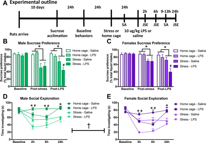
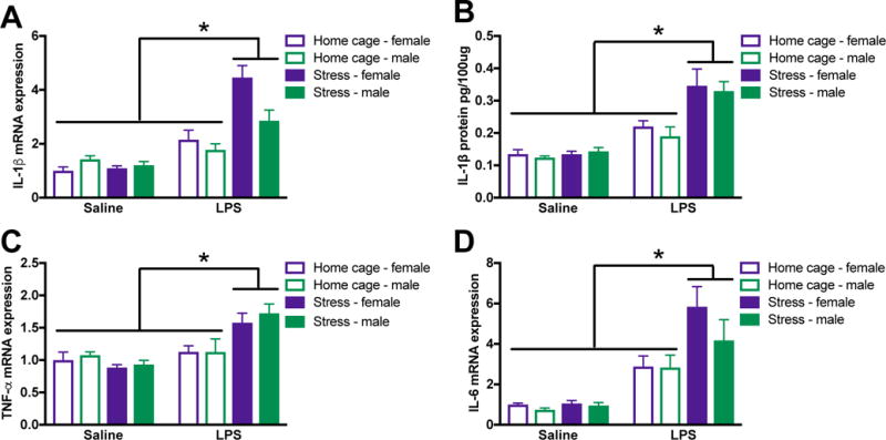
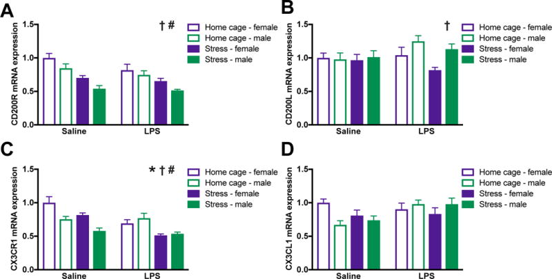
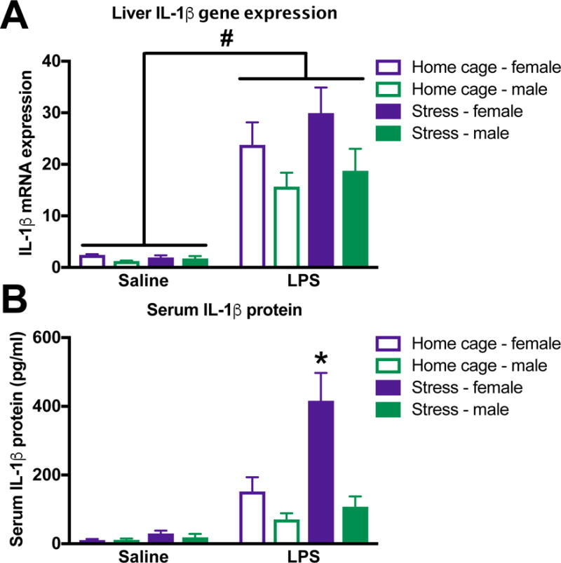
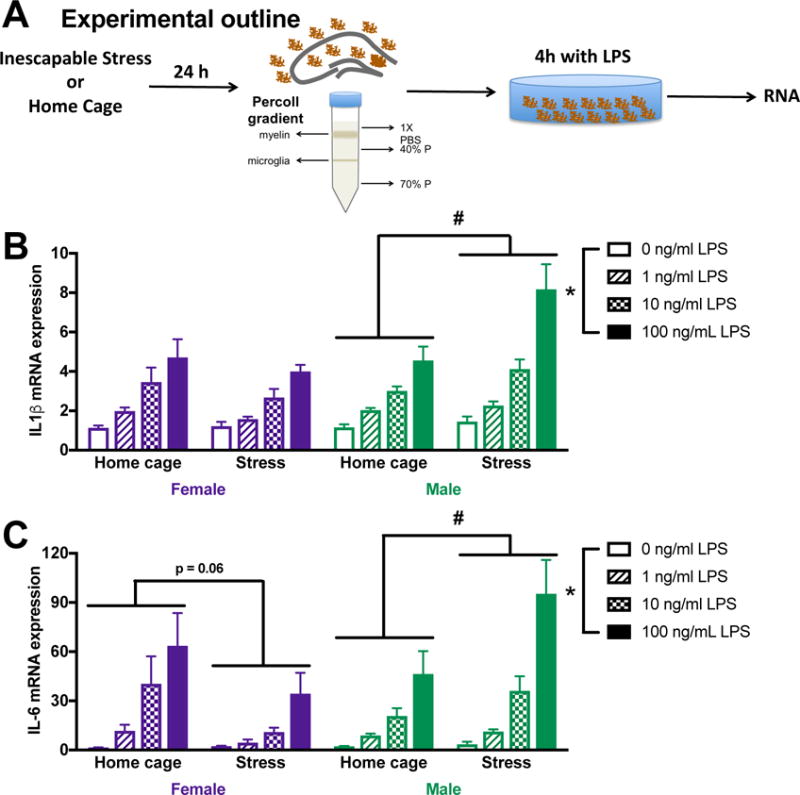
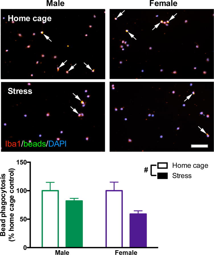
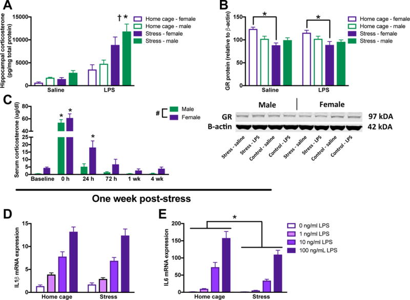
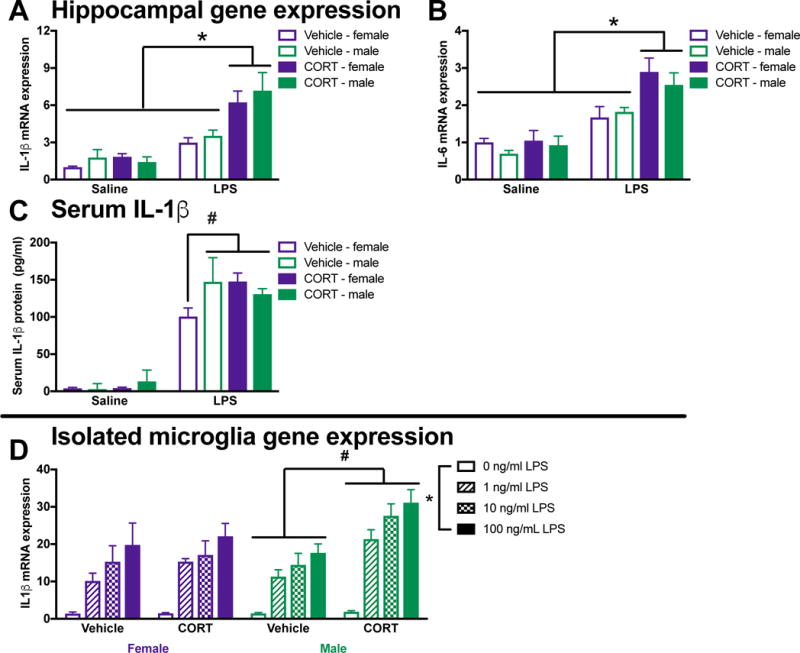
Exposure to stressors can enhance neuroinflammatory responses, and both stress and neuroinflammation are predisposing factors in the development of psychiatric disorders. Females suffer disproportionately more from several psychiatric disorders, yet stress-induced changes in neuroinflammation have primarily been studied in males. Here we tested whether exposure to inescapable tail shock sensitizes or 'primes' neuroinflammatory responses in male and female rats. At 24 h post-stress, male and female rats exposed to a peripheral immune challenge enhanced neuroinflammatory responses and exacerbated anxiety- and depressive-like behaviors. These changes are likely glucocorticoid dependent, as administering exogenous CORT, caused a similar primed inflammatory response in the hippocampus of male and female rats. Further, stress disinhibited anti-inflammatory signaling mechanisms (such as CD200R) in the hippocampus of male and female rats. In males, microglia are considered the likely cellular source mediating neuroinflammatory priming; stress increased cytokine expression in ex vivo male microglia. Conversely, microglia isolated from stressed or CORT treated females did not exhibit elevated cytokine responses. Microglia isolated from both stressed male and female rats reduced phagocytic activity; however, suggesting that microglia from both sexes experience stress-induced functional impairments. Finally, an immune challenge following either stress or CORT in females, but not males, increased peripheral inflammation (serum IL-1β). These novel data suggest that although males and females both enhance stress-induced neuroinflammatory and behavioral responses to an immune challenge, this priming may occur through distinct, sex-specific mechanisms.

Keywords: Glucocorticoids; Microglia; Neuroimmune; Sex differences; Sickness behavior; Stressors.


Conflict of interest statement

Conflict of interest: The authors declare no competing financial interests.

Figures

Figure 1
Figure 1. Sickness behavior is increased by prior stress in male and female rats
(A) Male and female rats underwent inescapable stress (100 trials of tailshock) or remained in the home cage. 24 h later rats received a single IP injection of 10 ug/kg LPS or saline (vehicle-control) and then underwent behavioral testing (see methods for additional details on timing). Male and female rats are graphed separately to simplify interpretation but sex was analyzed as a variable in both the sucrose preference and social exploration tests. (B) Male and (C) female rats demonstrated reduced sucrose preference following stress. Furthermore, prior stress exaggerated the LPS induced reduction in sucrose preference in both male and female rats. Juvenile social exploration was reduced by both stress and LPS in (D) male and (E) female rats. Furthermore, female rats had lower levels of social investigation as compared to male rats. Sucrose preference results were analyzed for each day using a 2 × 2 × 2 ANOVA with sex, stress, and immune challenge as the between subjects factors (n = 6 per group with a total of 48 rats). Results from the juvenile social exploration test were analyzed using 2 × 2 × 2 repeated measures ANOVAs with sex, stress, and immune challenge as the between subject factors and time as the within subjects factor. Data are expressed as mean ± SEM. main effect of sex, *main effect of stress, #main effect of LPS, p < 0.05 in all cases. Abbreviations: JSE (juvenile social investigation), SA (sucrose anhedonia), LPS (lipopolysaccharide).
Figure 2
Figure 2. Male and female rats exhibit comparable primed hippocampal cytokine responses post-stress
Male and female rats underwent inescapable stress or remained in the home cage. 24 h later rats received a single IP injection of 10 ug/kg LPS or saline and tissue was collected 3 h later. (A) Stress potentiated LPS-induced IL-1β mRNA elevations in the hippocampus of male and female rats. (B) IL-1β protein was similarly regulated in the hippocampus of male and female rats. Additional pro-inflammatory cytokine mRNA including (C) TNFα and (D) IL-6 were elevated by stress followed by LPS. Results were analyzed using 2 × 2 × 2 ANOVAs with sex, stress, and immune challenge as the between subjects factors (n = 6 – 8 per group with a total of 56 rats). Data are expressed as mean ± SEM. *simple effect of stress, p < 0.05 in all cases.
Figure 3
Figure 3. Male and female rats exhibited comparable down-regulation of anti-inflammatory pathway genes following stress
Male and female rats underwent inescapable stress or remained in the home cage. 24 h later rats received a single IP injection of 10 ug/kg LPS or saline and tissue was collected 3 h later. (A) CD200R mRNA was lower in the hippocampus of male as compared to female rats and was suppressed by stress in both sexes. (B) CD200L was increased in the hippocampus of male as compared to female rats and unaffected by and LPS stress. (C) CX3CR1 was reduced in the hippocampus of male as compared to female rats and suppressed by stress and LPS. (D) In contrast, mRNA expression of the CX3CL1 was unaffected by sex, stress, or LPS. Results were analyzed using 2 × 2 × 2 ANOVAs with sex, stress, and immune challenge as the between subjects factors (n = 6 – 8 per group with a total of 56 rats). Data are expressed as mean ± SEM. main effect of sex, *main effect of LPS, #main effect of stress, p < 0.05 in all cases.
Figure 4
Figure 4. Peripheral cytokine responses are potentiated in female but not male rats in response to stress and LPS
Female and male rats underwent inescapable stress or remained in the home cage. 24 h later rats received a single IP injection of 10 ug/kg LPS or saline and blood and tissue were collected 3 h later. (A) IL-1β mRNA expression in liver was increased by LPS but not stress in both female and male rats. (B) Serum IL-1β concentrations were increased by exposure to stress and LPS in female rats. In contrast, serum IL-1β was not affected by stress exposure in male rats. Results were analyzed using 2 × 2 × 2 ANOVAs with sex, stress, and immune challenge as the between subjects factors (n = 6 – 8 per group). Data are expressed as mean ± SEM. #main effect of LPS, *simple effect of stress, p < 0.05 in all cases.
Figure 5
Figure 5. Microglia are primed by exposure to stress in male but not female rats
(A) Male and female rats underwent inescapable stress or remained in the home cage. 24 h later microglia were isolated from the hippocampus using a percoll density gradient. Cells were plated (10,000 cell per well) with LPS for 4 h prior to isolating mRNA. (B) Prior stress exposure potentiated IL-1β mRNA responses to LPS in microglia isolated from male rats. In contrast, IL-1β mRNA expression was unaffected by prior stress in female microglia. (C) Similarly, IL-6 mRNA expression was potentiated by stress exposure in males, which trends toward an overall suppression in microglia isolated from the female hippocampus. Results were analyzed using 2 × 2 × 4 ANOVAs with sex, stress, and LPS dose as the independent variables (n = 4 per group and experiment replicated in an additional cohort of 32 female rats [supplemental Fig 1]). Data are expressed as mean ± SEM. *main effect of LPS, #stress X LPS interaction, p < 0.05 in all cases.
Figure 6
Figure 6. Stress reduces phagocytic capacity in microglia isolated from male and female rats
Male and female rats underwent inescapable stress or remained in the home cage. 24 h later microglia were isolated from the hippocampus using a percoll density gradient. Microglia were plated (5 × 10⁴ cells per well) with a latex bead solution (1:100 concentration). Microglia were identified with Iba1 (red), beads were conjugated to a green fluorophore, and nuclei were identified using DAPI (blue). Arrows highlight microglia that phagocytosed latex beads (yellow represents colocalization of red microglia and green beads). Stress caused fewer microglia to phagocytose the beads. Results were analyzed using a 2 × 2 ANOVA with sex and stress as the independent variables (n = 6 per group with a total of 24 rats; 20 images were analyzed per rat and averaged). Data are expressed as mean ± SEM. #main effect of stress, p < 0.05. Scale bar: 50 mm.
Figure 7
Figure 7. Some aspects of the glucocorticoid response are differentially modulated in male and female rats, but normalizing CORT concentrations does not result in priming in female microglia
(A) CORT responses are elevated by both prior stress and LPS in the hippocampus of male and female rats. (B) However, while glucocorticoid receptor concentrations are unaffected by stress in the hippocampus of male rats, females show downregulated glucocorticoid receptor expression in the hippocampus following stress. A representative blot is included below the graph (note: groups are represented in a different order in the blot than the figure). (C) The peripheral serum CORT response is elevated in females compared to males and CORT is increased for a longer duration of time following stress in females. Microglia isolated from female rats one week following stress when CORT concentrations have returned to baseline do not exhibit primed (D) IL-1β or (E) IL-6 mRNA expression. Panels A&B were analyzed using 2 × 2 × 2 ANOVAs with sex, stress, and immune challenge as the independent variables (n = 6 – 8 per group). Panel C was analyzed using a 2-way repeated measures ANOVA with sex as the between subjects factor and time as the within subject factor (n = 6 per group). Panels D&E were analyzed using 2 × 4 ANOVAs with stress and dose of LPS as the independent variables (n = 4 per group). Data are expressed as mean ± SEM. *main effect of stress, #main effect of sex, main effect of LPS, p < 0.05 in all cases.
Figure 8
Figure 8. Prior CORT treatment induces neuroinflammatory priming in vivo in male and female rats but does not prime female microglia
Male and female rats were injected with a single subcutaneous dose of CORT or vehicle (propylene glycol). 24 h later rats received 10 ug/kg IP LPS. Serum and hippocampi were collected after 3 h to evaluate cytokine expression. Prior CORT treatment potentiated LPS induced hippocampal IL-1β mRNA expression in (A) male and female rats. (B) Hippocampal IL-6 expression was similarly potentiated by prior CORT treatment in male and female rats. (C) Serum IL-1β was induced by an LPS injection in male rats but only potentiated by prior CORT treatment in females. In a separate cohort of rats, microglia were isolated 24 h following CORT or vehicle treatment. (D) Ex vivo treatment with LPS produced exaggerated IL-1β mRNA responses in male but not female rats that had received prior CORT treatment. Panels A – C were analyzed using 2 × 2 × 2 ANOVAs with sex, CORT, and LPS as the independent variables (n = 4 per group with a total of 32 rats). Panel D was analyzed using 2 × 2 × 4 ANOVAs with sex, CORT, and LPS dose as the independent variables (n = 4 per group with a total of 16 rats). Data are expressed as mean ± SEM. *main effect of LPS, #interaction of CORT and LPS (G), p < 0.05 in all cases.

Comment in

References

    1. Arakawa K, Arakawa H, Hueston CM, Deak T. Effects of the estrous cycle and ovarian hormones on central expression of interleukin-1 evoked by stress in female rats. Neuroendocrinology. 2014;100:162–177. - PubMed
    1. Barrientos RM, V, Thompson M, Kitt MM, Amat J, Hale MW, Frank MG, Crysdale NY, Stamper CE, Hennessey PA, Watkins LR, Spencer RL, Lowry CA, Maier SF. Greater glucocorticoid receptor activation in hippocampus of aged rats sensitizes microglia. Neurobiol Aging. 2015;36:1483–1495. - PMC - PubMed
    1. Bekhbat M, Neigh GN. Sex differences in the neuro-immune consequences of stress: Focus on depression and anxiety. Brain Behav Immun 2017 - PMC - PubMed
    1. Biber K, Neumann H, Inoue K, Boddeke HW. Neuronal ‘On’ and ‘Off’ signals control microglia. Trends Neurosci. 2007;30:596–602. - PubMed
    1. Bourke CH, Harrell CS, Neigh GN. Stress-induced sex differences: adaptations mediated by the glucocorticoid receptor. Horm Behav. 2012;62:210–218. - PMC - PubMed

Publication types